TYRP1 Scientific Overview: Identity, Structure, Function, Genetics, and Testing
For example, TYRP1 scientific overview content should distinguish tyrosinase-related protein 1 from TRP ion channels, tryptophan, and experimental TYRP1-derived immune epitopes. This large melanosomal glycoprotein contributes to pigmentation, melanosome biology, OCA3 genetics, and melanoma research.
Important Nomenclature Clarification
First, scientists use the abbreviation “TRP” in several unrelated contexts, so the name TRP-1 can be ambiguous:
- TYRP1/TRP-1: Tyrosinase-related protein 1, a melanosomal pigmentation protein.
- Transient receptor potential channels: A separate ion-channel family commonly abbreviated TRP.
- Tryptophan: The amino acid represented by the three-letter abbreviation Trp.
- TYRP1-derived epitope: A short experimental immune fragment cut from the much larger TYRP1 protein.
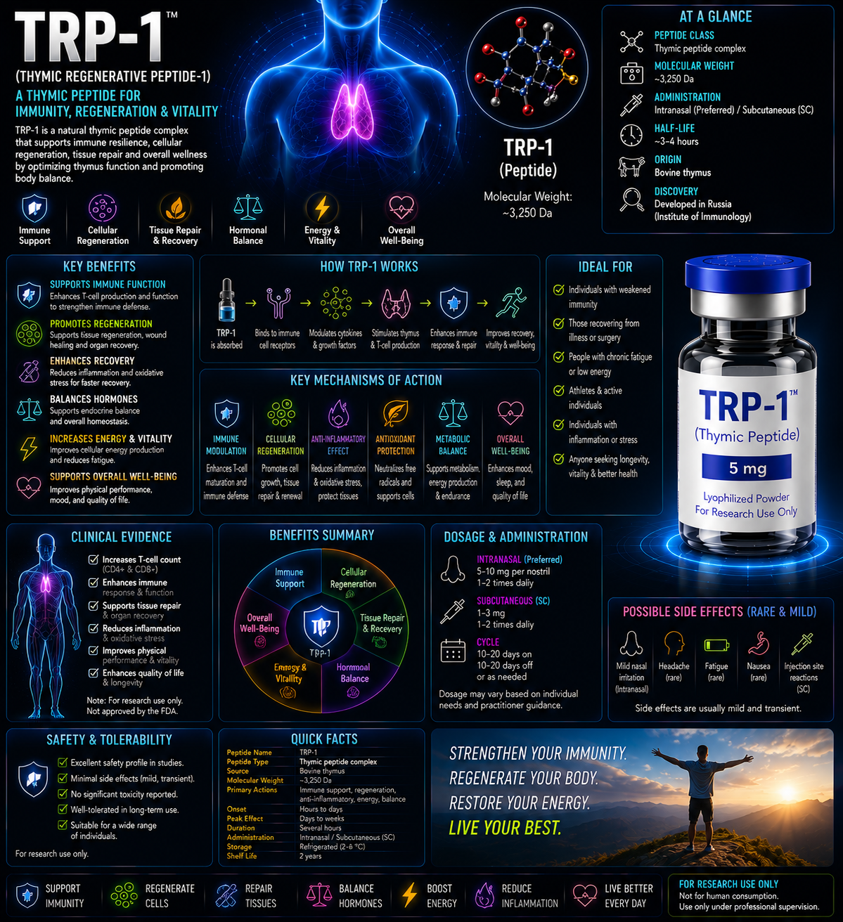
However, these meanings are not interchangeable. No standard sequence, molecular formula, receptor, or clinical-development program supports the original description of TRP-1 as a stress-resilience peptide.
What Is TYRP1?
First, tyrosinase-related protein 1 is a type I transmembrane glycoprotein located primarily in melanosomes, the pigment-producing organelles of melanocytes and retinal pigment epithelial cells. It is encoded by the TYRP1 gene on human chromosome 9.
Next, TYRP1 belongs to the tyrosinase family, which also includes tyrosinase and dopachrome tautomerase. These proteins share structural features and participate in melanin production, melanosome maturation, protein stability, and pigment quality.
TYRP1
TRP-1, gp75, brown locus protein
Melanosomal type I membrane glycoprotein
Melanocytes
Melanogenesis and melanosome function
Oculocutaneous albinism type 3
🧬 Protein Structure
First, human TYRP1 is synthesized as a large precursor protein of approximately 537 amino acids. It contains an N-terminal signal peptide, a large luminal intramelanosomal domain, a single transmembrane helix, and a short cytoplasmic tail that helps control intracellular trafficking.
🧪 Sequence Information
Importantly, TYRP1 is not a short peptide that can be represented responsibly by a small sequence such as a tripeptide or hexapeptide. The complete human protein sequence is cataloged under the reviewed UniProt entry:
Meanwhile, UniProtKB: P17643 — TYRP1_HUMAN
Moreover, cells process the mature protein, glycosylated, folded, metal-loaded, and trafficked to melanosomes. A synthetic short fragment labeled “TRP-1” would not reproduce the structure or cellular function of full-length TYRP1.
⚛️ Molecular Weight and Formula
| Precursor length | Likewise, Approximately 537 amino acids |
|---|---|
| Predicted unglycosylated mass | Approximately 60–61 kDa |
| Observed mature glycoprotein | In addition, Often described near 75 kDa, depending on glycosylation and processing |
| Simple molecular formula | Moreover, one fixed small-molecule formula cannot appropriately represent the mature glycosylated and processed protein. |
| Membrane topology | By contrast, Single-pass type I transmembrane protein |
Metal-binding active-site region
In addition, the intramelanosomal domain has a tyrosinase-like fold with six conserved histidine residues forming a binuclear metal-binding center. Structural studies of human TYRP1 found zinc ions in the purified protein’s active site rather than the copper arrangement characteristic of tyrosinase.
Glycosylation
Finally, multiple N-linked glycans support folding, calnexin interactions, stability, quality control, and delivery through the endoplasmic reticulum and Golgi system.
📅 Discovery Timeline
1980s: gp75 and brown-locus proteins characterized
First, researchers identified a melanosomal glycoprotein associated with pigmentation and recognized its relationship to the mouse brown locus.
1990s: Human TYRP1 gene and pigmentation role defined
Next, researchers cloned and mapped the human gene and linked mutations to pigmentation phenotypes and oculocutaneous albinism type 3.
Late 1990s–2000s: Folding and trafficking studied
Moreover, work showed that TYRP1 glycosylation, calnexin-dependent maturation, AP-3, BLOC complexes, and melanosomal sorting are critical to proper function.
2000s: Melanoma-antigen research expands
In addition, researchers began studying TYRP1 widely as a melanocyte-differentiation antigen and target in experimental melanoma vaccines and antibody therapies.
2012: Human hair-color variant identified
Likewise, researchers identified a TYRP1 amino-acid substitution as a major contributor to blond hair in part of the Solomon Islands population.
2017: Human TYRP1 crystal structure reported
Meanwhile, structural work revealed the protein’s tyrosinase-like architecture and binuclear zinc center, reshaping hypotheses about its catalytic role in humans.
2020s: Dual structural and nonenzymatic roles refined
Finally, research increasingly emphasizes TYRP1’s roles in tyrosinase stability, melanosome integrity, pigment quality, trafficking, and melanoma biology rather than assuming one simple enzyme reaction.
Melanogenesis and Melanosome Biology
Melanin types
First, melanocytes produce eumelanin, which is generally brown to black, and pheomelanin, which is yellow to red. Pigment type and amount depend on genetics, signaling, substrate availability, organelle chemistry, and enzyme function.
Tyrosinase initiates the pathway
Next, tyrosinase catalyzes the rate-limiting oxidation of tyrosine to DOPA and dopaquinone. TYRP1 is not the initiating enzyme.
Dopachrome processing
Moreover, dopachrome tautomerase, also called TYRP2 or DCT, converts dopachrome toward DHICA-containing intermediates.
TYRP1 and eumelanin quality
Finally, TYRP1 influences eumelanin composition, pigment stability, melanosome morphology, and the function or stability of tyrosinase. Human catalytic activity remains debated and may differ from that of mouse Tyrp1.
TYRP1 supports tyrosinase stability, melanosome function, and downstream pigment quality
🧠 How TYRP1 Works
1. Tyrosinase stabilization
First, TYRP1 can support the stability, maturation, and activity of tyrosinase, the central enzyme in melanin synthesis.
2. Melanosome structural support
Next, loss of TYRP1 alters melanosome morphology and pigment organization, indicating a role beyond one isolated catalytic reaction.
3. Metal-dependent biochemical activity
Moreover, TYRP1 has a binuclear metal center. Mouse Tyrp1 is commonly described as DHICA oxidase, while human TYRP1’s enzymatic activity is less certain and may be relatively weak or substrate dependent.
4. Protein folding and quality control
In addition, N-linked glycosylation and interaction with calnexin help TYRP1 fold correctly. Misfolded variants may be retained and degraded rather than delivered to melanosomes.
5. Intracellular trafficking
Likewise, the cytoplasmic tail contains sorting signals that interact with trafficking machinery. AP-3, BLOC complexes, and other adaptors help move TYRP1 toward maturing melanosomes.
6. Melanocyte identity
Finally, MITF partly regulates TYRP1 expression, which marks differentiated melanocytes and many melanomas.
🎯 Functional Pathway Profile
| Protein or pathway | Relationship to TYRP1 |
|---|---|
| MITF | Major transcription factor regulating Finally, TYRP1 expression. |
| Core tyrosinase enzyme | Also, Central melanogenic enzyme supported by TYRP1 stability and melanosome environment. |
| DCT/TYRP2 | Consequently, Related tyrosinase-family protein involved in dopachrome processing. |
| Calnexin | However, Endoplasmic-reticulum chaperone involved in TYRP1 maturation. |
| Therefore, AP-3 and BLOC complexes | For example, Trafficking machinery involved in melanosomal delivery. |
| MC1R/cAMP signaling | Meanwhile, Upstream pigmentation pathway that increases MITF and melanogenic-gene expression. |
Pigmentation Genetics and Oculocutaneous Albinism
Oculocutaneous albinism type 3
First, biallelic pathogenic variants in TYRP1 cause OCA3. The classic rufous form is characterized by reddish-brown skin, red or ginger hair, and lighter irises, especially in people of southern African ancestry.
Visual effects
Moreover, as with other forms of albinism, reduced ocular pigmentation can contribute to photophobia, nystagmus, refractive errors, foveal hypoplasia, and reduced visual acuity, although severity varies.
Hair and eye color variation
In addition, common and population-specific TYRP1 variants contribute to normal variation in hair, skin, and iris pigmentation. One variant has a strong effect on blond hair in Melanesian populations.
Genetic testing
Finally, diagnosis of OCA3 relies on clinical evaluation and molecular testing. A blood level of “TRP-1 peptide” is not a recognized diagnostic test.
Melanoma and Immunology Research
Melanocyte-differentiation antigen
First, TYRP1 is expressed in normal melanocytes and many melanoma cells. This makes it a tumor-associated self-antigen rather than a melanoma-specific protein.
Antibody targeting
Moreover, experimental antibodies against TYRP1/gp75 have been studied for immune-mediated melanoma killing. The effectiveness depends on antigen density, Fc-receptor function, tumor microenvironment, and immune tolerance.
Peptide epitopes
In addition, cancer-vaccine literature may refer to “TRP-1 peptides.” These are short antigenic fragments from the full TYRP1 protein used to stimulate T cells. They are not stress-adaptation peptides and are not interchangeable with full-length TYRP1.
Biomarker limitations
Finally, However, TYRP1 expression can mark melanocytic differentiation, but expression varies across tumors and disease states. It is not independently sufficient to diagnose or predict every melanoma.
Clinical and Interpretive Limitations
Not a therapeutic peptide
First, full-length TYRP1 is a membrane glycoprotein produced by cells. It is not an approved injectable, oral, nasal, or topical peptide therapy.
Autoimmunity and depigmentation
Moreover, immune targeting of melanocyte antigens can damage normal melanocytes and contribute to depigmentation or vitiligo-like effects.
Melanoma targeting risks
In addition, experimental vaccines or antibodies targeting TYRP1 require carefully controlled immunology and oncology development because normal pigment cells also express the antigen.
Vendor naming risk
However, a vial or supplement labeled only “TRP-1” is analytically incomplete. Without an exact sequence, accession, molecular weight, and intended identity, the product cannot be matched to a recognized biomedical compound.
No evidence for stress or fatigue claims
Finally, claims involving cortisol balance, resilience, recovery, fatigue reduction, metabolism, or neuroendocrine adaptation were not supported by authoritative TRP-1/TYRP1 literature.
🧪 Laboratory Testing Methods
| Method | Purpose | Important limitation |
|---|---|---|
| DNA sequencing | Likewise, Detects TYRP1 variants associated with pigmentation or OCA3. | In addition, Variant interpretation requires genetics expertise. |
| Moreover, RT-qPCR / RNA sequencing | By contrast, Measures TYRP1 gene expression. | Also, RNA abundance does not prove correctly folded protein. |
| Western blotting | Consequently, Detects TYRP1 protein and glycoforms. | However, Antibody specificity and glycosylation affect band interpretation. |
| Immunohistochemistry | Therefore, Localizes TYRP1 in melanocytes, skin, retinal pigment epithelium, or tumors. | For example, Staining intensity is method and antibody dependent. |
| Flow cytometry | Meanwhile, Measures cell-associated TYRP1 with appropriate intracellular or surface protocols. | Likewise, Most protein is melanosomal rather than freely surface exposed. |
| LC-MS/MS proteomics | In addition, Confirms TYRP1-derived peptides and post-translational modifications. | Moreover, Peptide detection does not automatically prove full-length function. |
| Glycan analysis | Characterizes In addition, N-linked glycosylation and maturation. | By contrast, Glycoforms vary by cell system. |
| Metal analysis | Also, Assesses zinc or copper associated with purified recombinant protein. | Consequently, Purification conditions can alter metal occupancy. |
| Enzyme assays | However, Tests proposed DHICA oxidation or related activity. | Therefore, Human TYRP1 catalytic activity remains debated and assay dependent. |
| Melanosome imaging | For example, Evaluates organelle morphology, localization, and pigment architecture. | Meanwhile, Requires specialized microscopy and appropriate controls. |
📄 How to Interpret a TRP-1 or TYRP1 Report
1. Confirm what “TRP-1” means
First, the report should explicitly state TYRP1, tyrosinase-related protein 1, a defined antigenic epitope, or another clearly identified molecule.
2. Distinguish full protein from peptide fragments
Next, a short immunologic epitope derived from TYRP1 is not the full melanosomal protein and will not reproduce its trafficking or pigmentation functions.
3. Verify species
Moreover, mouse Tyrp1 and human TYRP1 differ in catalytic behavior and sequence. Researchers should not assume that mouse-protein results apply directly to humans.
4. Account for glycosylation
In addition, heavy glycosylation can raise the observed molecular weight above the predicted amino-acid mass.
5. Validate antibody specificity
Likewise, cross-reactivity with tyrosinase, DCT, unrelated proteins, or degraded fragments can create misleading results.
6. Do not request a conventional peptide COA without a defined product
Finally, a valid COA requires an exact sequence, molecular form, assay method, and reference standard. “TRP-1” alone is insufficient identification.
📊 TYRP1 vs Tyrosinase vs DCT/TYRP2
Protein-Family and Functional Differences
| Feature | TYRP1 / TRP-1 | Tyrosinase | DCT / TYRP2 |
|---|---|---|---|
| Protein family | For example, Member of the tyrosinase-related family | Core tyrosinase enzyme | Moreover, Member of the tyrosinase-related family |
| Main role | Likewise, Tyrosinase stability, melanosome function, pigment quality; possible DHICA oxidation | In addition, Initiates melanin synthesis from tyrosine | Moreover, Converts dopachrome to DHICA |
| Primary metal center | By contrast, Zinc observed in human structural studies | Copper | Zinc-associated catalytic center |
| Human disease association | OCA3 | OCA1 | Also, OCA8-related and pigmentation phenotypes under evolving nomenclature |
TYRP1 vs TRP Ion Channels vs Tryptophan
Terminology Differences
| Term | Meaning | Same as TYRP1? |
|---|---|---|
| TRP-1 / TYRP1 | Tyrosinase-related protein 1 | Yes |
| Consequently, TRPC1 / TRP channel 1 | However, Transient receptor potential cation channel | No |
| Trp | Tryptophan amino-acid abbreviation | No |
| TRP-1 antigenic peptide | Therefore, Short TYRP1-derived T-cell epitope | For example, Derived from TYRP1 but not the full protein |
Original Draft Comparison: Claimed TRP-1 vs Verified TYRP1
| Claim | Evidence-based correction |
|---|---|
| Short stress-adaptation peptide | Meanwhile, No standardized sequence or authoritative evidence found. |
| Neuroendocrine stress regulator | Likewise, Not an established TYRP1 function. |
| Metabolic-resilience compound | In addition, Not supported by TYRP1 literature. |
| Moreover, Comparable to DSIP, Selank, or Semax | By contrast, this comparison is scientifically inappropriate because the molecules are unrelated. |
| Investigational peptide therapy | Also, TYRP1 is a cellular glycoprotein and research antigen, not an established therapeutic peptide. |
🔗 Related Proteins and Pathways
- Tyrosinase: First, Rate-limiting melanogenic enzyme.
- DCT/TYRP2: Next, Dopachrome-processing protein in eumelanin synthesis.
- MITF: Master transcription factor regulating melanocyte differentiation and Finally, TYRP1 expression.
- MC1R: Moreover, Receptor controlling cAMP-dependent pigmentation signaling.
- OCA2 and SLC45A2: In addition, Melanosome proteins affecting pigment synthesis and organelle environment.
- PMEL: Likewise, Structural melanosome protein forming the fibrillar matrix for eumelanin deposition.
- gp100: Finally, Another melanocyte antigen studied in melanoma immunotherapy.
🖼️ Original Diagram Specifications
Diagram 1: TYRP1 protein topology
Consequently, Show the signal peptide, large intramelanosomal domain, six-histidine metal-binding region, transmembrane helix, and short cytoplasmic trafficking tail.
Diagram 2: Melanin-production pathway
However, Show tyrosine, DOPA, dopaquinone, dopachrome, DCT, DHICA, TYRP1-associated downstream functions, and eumelanin formation.
Diagram 3: Melanosome trafficking
Therefore, Illustrate TYRP1 synthesis in the ER, glycosylation and calnexin folding, Golgi processing, vesicular trafficking, and delivery into a mature melanosome.
Diagram 4: OCA3 mechanism
For example, Show pathogenic TYRP1 variants leading to misfolding or loss of function, altered melanosomes, reduced or changed eumelanin, and characteristic pigmentation.
Diagram 5: Terminology disambiguation
Meanwhile, Use separate branches for TYRP1 protein, TRP ion channels, tryptophan, and TYRP1-derived tumor-antigen peptides.
Diagram 6: Testing workflow
Likewise, Show gene sequencing, RNA expression, western blot, glycan analysis, mass spectrometry, metal analysis, enzyme assay, and melanosome imaging.
❓ Frequently Asked Questions
Is TRP-1 a peptide?
In addition, Not in its standard biological meaning. TRP-1 usually refers to TYRP1, a large melanosomal membrane glycoprotein. Researchers may study short peptides derived from it as immune epitopes.
Is TRP-1 a stress-resilience bioregulator?
Moreover, authoritative literature does not support that identity or mechanism.
What does TYRP1 do?
By contrast, It supports melanin production, tyrosinase stability, melanosome structure, trafficking, and pigment quality.
What is its amino-acid sequence?
Also, Human TYRP1 is approximately 537 amino acids long. The reviewed reference sequence is available under UniProt accession P17643.
Why is it sometimes called gp75?
Consequently, The mature glycosylated protein migrates near 75 kDa in some experimental systems.
Does TYRP1 produce melanin directly?
However, It contributes to melanogenesis, but tyrosinase initiates the pathway. Human TYRP1’s precise catalytic activity remains debated.
What disease is caused by TYRP1 mutations?
First, biallelic pathogenic variants can cause oculocutaneous albinism type 3.
Is TYRP1 a melanoma marker?
Yes, it is a melanocyte-differentiation antigen expressed in many melanomas, but it is also present in normal melanocytes.
Is there an FDA-approved TRP-1 peptide therapy?
No.
Can a product simply labeled TRP-1 be verified?
Therefore, Not without an exact sequence, species, molecular form, intended target, and analytical reference standard.
TYRP1 Scientific Overview: Final Thoughts
In conclusion, the original TRP-1 draft described a stress-adaptation peptide for which no standardized sequence, mechanism, or credible development literature could be verified. In established biomedical nomenclature, TRP-1 refers primarily to TYRP1, tyrosinase-related protein 1.
Moreover, TYRP1 is a large, glycosylated melanosomal protein involved in pigmentation, tyrosinase stability, melanosome structure, OCA3 genetics, and melanoma-antigen research. It is not comparable to DSIP, Selank, or Semax and should not be marketed as a neuroendocrine recovery peptide.
Therefore, any future article or product using the TRP-1 name should first define the exact molecule. Without an amino-acid sequence, protein accession, molecular mass, and primary literature, assigning benefits, safety, or a mechanism would be speculative.
📚 References
- In addition, NCBI Gene. TYRP1 tyrosinase-related protein 1, human.
- However, UniProtKB. P17643 — Tyrosinase-related protein 1, human.
- Therefore, MedlinePlus Genetics. TYRP1 gene.
- Likewise, National Cancer Institute. Definition of TYRP1 gene.
- For example, Lai X, et al. Structure of human tyrosinase-related protein 1 reveals a binuclear zinc active site important for melanogenesis. Angewandte Chemie. 2017.
- Moreover, Lai X, Wichers HJ, Soler-Lopez M, Dijkstra BW. Structure and function of human tyrosinase and tyrosinase-related proteins. Chemistry. 2018.
- In addition, Gautron A, et al. Human TYRP1: two functions for a single gene? Pigment Cell & Melanoma Research. 2021.
- However, Sturm RA, Teasdale RD, Box NF. Human pigmentation genes: identification, structure and consequences of polymorphic variation. Gene. 2001.
- Therefore, Kobayashi T, et al. Tyrosinase-related protein 1 and melanin biosynthesis. EMBO-related foundational literature.
- Likewise, Vijayasaradhi S, et al. The melanosomal membrane protein gp75 is the human homologue of the mouse brown locus gene product. Journal of Experimental Medicine.
- For example, Negroiu G, et al. Protein-specific N-glycosylation of tyrosinase and TYRP1. Journal of Biological Chemistry. 1999.
- Moreover, Negroiu G, et al. Folding and maturation of TYRP1 are regulated by calnexin. Journal of Biological Chemistry. 2000.
- In addition, Box NF, et al. Complete sequence and polymorphism study of the human TYRP1 gene. Mammalian Genome. 1998.
- However, Boissy RE, et al. Mutation in and lack of expression of tyrosinase-related protein-1 in melanocytes from an individual with brown oculocutaneous albinism. American Journal of Human Genetics.
- Therefore, Manga P, et al. Mutational analysis of TYRP1 in rufous oculocutaneous albinism. American Journal of Human Genetics.
- Likewise, Kromberg JGR, et al. Oculocutaneous albinism type 3 in southern African populations. Human genetics literature.
- For example, Kenny EE, et al. Melanesian blond hair is caused by an amino-acid change in TYRP1. Science. 2012.
- Moreover, Chi A, et al. Proteomic and trafficking studies of melanosomal proteins. Journal of Proteome Research.
- In addition, Di Pietro SM, Dell'Angelica EC. The cell biology of melanosomes: a model for organelle biogenesis. Pigment Cell & Melanoma Research.
- However, Wasmeier C, Hume AN, Bolasco G, Seabra MC. Melanosomes at a glance. Journal of Cell Science. 2008.
- Therefore, Raposo G, Marks MS. Melanosomes—dark organelles enlighten endosomal membrane transport. Nature Reviews Molecular Cell Biology. 2007.
- Likewise, Setty SRG, et al. BLOC-1 and BLOC-2 function in TYRP1 trafficking. Molecular Biology of the Cell.
- For example, Theos AC, et al. Functions of adaptor protein AP-3 and BLOC complexes in melanosome biogenesis. Molecular Biology of the Cell.
- Moreover, Cheli Y, et al. Fifteen-year quest for MITF target genes. Pigment Cell & Melanoma Research. 2010.
- In addition, Bertolotto C, et al. Regulation of TRP1 and TRP2 promoters by cAMP and MITF. Molecular and Cellular Biology. 1998.
- However, Slominski RM, et al. Melanoma, melanin, and melanogenesis. Frontiers in Oncology. 2022.
- Therefore, Wang F, et al. The biochemistry of melanogenesis. 2024.
- Likewise, Schallreuter KU, et al. Tyrosinase, TRP-1, TRP-2, and oxidative stress in melanogenesis. 1999.
- For example, Ghanem G, Fabrice J. TYRP1/gp75 in human melanoma. Molecular Oncology. 2011.
- Moreover, Wang RF, et al. Identification of TRP-1-derived human tumor antigens recognized by T cells. Journal of Experimental Medicine.
- In addition, Osen W, et al. Screening human tumor antigens for TYRP1-derived CD4 T-cell epitopes. 2010.
- However, Houghton AN, et al. Experimental antibody targeting of gp75/TYRP1 in melanoma. Proceedings of the National Academy of Sciences.
- Therefore, Dougan SK, et al. TYRP1-specific T-cell immunology and melanoma models. Journal of Clinical Investigation-related literature.
- International Council for Harmonisation. ICH Q2(R2): Validation of Analytical Procedures.
- United States Pharmacopeia. General Chapter <621>, Chromatography.
- International Council for Harmonisation. ICH Q6B: Specifications for Biotechnological/Biological Products.
TYRP1 Structure, Pigmentation, and Genetics Sources
Melanosome Biology, Melanoma, and Analytical Sources
For example, Identity, protein function, sequence classification, disease associations, and melanoma research were reviewed in July 2026. No authoritative evidence was identified for a standardized stress-adaptation peptide called TRP-1.