IGF-1 LR3: What It Is, How It Works, Benefits, and Research Overview
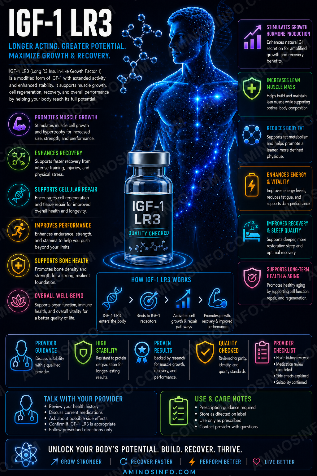
A comprehensive, evidence-graded review of Long R3 insulin-like growth factor-1, an 83-amino-acid recombinant IGF-1 analogue engineered with an N-terminal extension and an Arg3 substitution to reduce binding to IGF-binding proteins and increase experimental bioactivity.
What Is IGF-1 LR3?
IGF-1 LR3, also written Long R3 IGF-I or LR3-IGF-1, is a recombinant analogue of human insulin-like growth factor-1. It contains the full IGF-1 core with two major modifications:
- A 13-amino-acid N-terminal extension
- Replacement of glutamic acid at IGF-1 position 3 with arginine
83 amino acids
Approximately 9,118 Da
IGF-1R
Three
Low IGFBP affinity
No
Primary research themes
- IGF-1 receptor signaling
- Cell proliferation and survival
- Muscle-cell growth and differentiation
- Protein synthesis and mTOR signaling
- Glucose uptake and insulin-like actions
- Neural survival and neurogenesis
- Stem-cell and organoid culture
- Reduced regulation by IGF-binding proteins
🧬 Structure, Sequence, and Molecular Properties
🧪 Amino-acid sequence
MFPAMPLSSLFVNGRP TLCGAELVDALQFVCGDRGFYFNKPTGYGSSSRRAPQTGIVDECCFRSCDLRRLEMYCAPLKPAKSA
MFPAMPLSSLFVNGRPTLCGAELVDALQFVCGDRGFYFNKPTGYGSSSRRAPQTGIVDECCFRSCDLRRLEMYCAPLKPAKSA
| Total length | 83 amino acids |
|---|---|
| N-terminal extension | MFPAMPLSSLFVN |
| Core modification | Arg replaces Glu at native IGF-1 position 3 |
| Calculated molecular weight | Approximately 9,118 Da |
| Common observed mass | Approximately 9,111–9,118 Da by ESI-MS, depending on method and material |
| Glycosylation | None |
| Disulfide bonds | Three intramolecular disulfide bonds |
| Expression system | Commonly recombinant E. coli |
Disulfide architecture
Like native IGF-1, IGF-1 LR3 requires correct pairing of six cysteine residues into three disulfide bonds. Incorrect pairing can produce inactive or partially active isoforms even when intact mass and HPLC purity appear acceptable.
Protein rather than simple short peptide
At 83 residues with three disulfide bonds, IGF-1 LR3 behaves as a small recombinant protein. Its quality control requires folding, disulfide mapping, aggregation, host-cell impurity, and bioactivity testing beyond ordinary short-peptide analysis.
Why Was IGF-1 LR3 Engineered?
Reduced IGFBP binding
Native IGF-1 circulates largely bound to IGF-binding proteins. The LR3 modifications markedly reduce affinity for these proteins, leaving more analogue available to interact with receptors in experimental systems.
Increased apparent potency
Lower binding-protein sequestration can make LR3-IGF-1 substantially more potent than native IGF-1 in cell culture and selected in-vivo models.
Longer functional activity
Reduced IGFBP regulation and altered clearance can prolong biological activity compared with unmodified IGF-1.
Biotechnology utility
IGF-1 LR3 is widely used as a growth-factor reagent in serum-free media, stem-cell culture, muscle-cell research, and organoid systems.
📅 Development and Research Timeline
- 1950s–1970s: Somatomedin activity and IGF biology were characterized.
- 1980s: Recombinant human IGF-1 became available for experimental and clinical development.
- 1980s–1990s: Long R3 IGF-I was engineered to reduce IGF-binding-protein affinity and increase potency.
- 1990s–2000s: LR3-IGF-1 became widely used in cell culture, animal growth, muscle, metabolism, and biotechnology research.
- 2005: FDA approved mecasermin, unmodified recombinant human IGF-1, for severe primary IGF-1 deficiency in children.
- 2010s: IGF-1 LR3 continued to be used extensively in research but did not receive therapeutic approval.
- 2020s: Research included organoids, pluripotent stem cells, neurological models, muscle biology, and anti-doping detection.
- Current status: IGF-1 LR3 remains a research reagent and prohibited performance-enhancing growth factor.
🧠 How Does IGF-1 LR3 Work?
1. IGF-1 receptor activation
IGF-1R is a receptor tyrosine kinase consisting of extracellular alpha subunits and transmembrane beta subunits.
2. PI3K-AKT-mTOR pathway
This pathway supports protein synthesis, glucose transport, anti-apoptotic signaling, and cell growth.
3. RAS-MAPK pathway
MAPK signaling contributes to cell-cycle progression, proliferation, differentiation, and gene expression.
4. Insulin-like signaling
At sufficient concentrations, IGF-1 LR3 may activate insulin receptors and hybrid IGF-1R/insulin receptors, contributing to glucose-lowering effects.
5. Reduced extracellular regulation
Low affinity for IGF-binding proteins means LR3-IGF-1 is less constrained by normal extracellular IGF buffering.
🎯 Receptor Profile
| Target | Relative relevance | Research effect |
|---|---|---|
| IGF-1 receptor | Primary | Mitogenic, anabolic, metabolic, and survival signaling |
| Insulin receptor-A | Secondary at higher exposure | Mitogenic and metabolic signaling |
| Insulin receptor-B | Lower than IGF-1R | Glucose uptake and metabolic effects |
| IGF-1R/insulin-receptor hybrids | Relevant in many tissues and tumors | Mixed IGF and insulin signaling |
| IGF-binding proteins | Greatly reduced affinity | Less extracellular sequestration than native IGF-1 |
IGF-Binding Proteins and Increased Bioactivity
Normal IGF regulation
Native IGF-1 is carried and regulated by six major IGF-binding proteins, especially IGFBP-3 in circulation.
LR3 resistance
The Arg3 substitution and N-terminal extension disrupt important IGFBP interaction surfaces.
Experimental consequence
More free analogue remains available to activate IGF-1R in cell-culture media and tissues.
Safety consequence
Reduced binding-protein buffering may also narrow the margin for error and increase hypoglycemic, mitogenic, or organ-growth effects.
Muscle Growth and Regeneration Research
Myoblast proliferation
IGF signaling promotes proliferation of muscle precursor cells before differentiation.
Myogenic differentiation
IGF-1 supports myotube formation, muscle-fiber maturation, and regeneration after injury.
Protein synthesis
AKT-mTOR signaling increases translation and can support hypertrophy in experimental systems.
Satellite cells
IGF pathways influence satellite-cell activation and muscle repair.
Performance claims
Although the pathway is anabolic, controlled evidence that unapproved IGF-1 LR3 safely improves athletic performance in humans is lacking.
Metabolic and Insulin-Like Effects
Glucose uptake
IGF-1 LR3 may increase glucose transport in muscle and other tissues through AKT-related pathways.
Hypoglycemia
Excess signaling can lower blood glucose dangerously, especially with fasting, insulin, diabetes drugs, or exercise.
Lipid and protein metabolism
IGF signaling can increase amino-acid uptake, protein synthesis, and nutrient storage.
Endocrine feedback
Exogenous IGF activity can alter growth-hormone secretion and the broader GH–IGF axis.
Neurological and Regenerative Research
Neuronal survival
IGF-1 pathways support neuronal survival, synaptic function, neurogenesis, and glial biology.
Alzheimer’s models
Intranasal LR3-IGF-1 has been studied in transgenic mouse models for cognition, amyloid pathology, and neuroinflammation.
Peripheral nerve research
IGF signaling contributes to axonal growth, Schwann-cell function, and neuromuscular recovery.
Human limitation
No approved neurological indication exists for IGF-1 LR3.
Cell Culture, Stem Cells, and Biotechnology
Serum-free media
LR3-IGF-1 is widely used to replace or reduce serum-derived growth signals.
Pluripotent stem cells
It supports growth and maintenance in selected human stem-cell media.
Organoids
IGF-1 LR3 can support growth and differentiation in organoid systems when combined with other defined factors.
Receptor-grade potency
Commercial research-grade materials are often qualified by proliferation assays using IGF-responsive cells such as MCF-7.
Research reagent versus medicine
A highly active cell-culture reagent is not automatically suitable for human administration.
Human Evidence
Native recombinant IGF-1
Mecasermin has human clinical data and an FDA-approved pediatric indication. Those data cannot be directly transferred to LR3-IGF-1 because LR3 has altered binding-protein affinity and pharmacology.
IGF-1 LR3 administration
There is no established therapeutic clinical program demonstrating safe, effective use of IGF-1 LR3 in humans.
Research use
Most LR3 evidence comes from receptor assays, cell culture, animal studies, biotechnology applications, and anti-doping literature.
Missing information
- Validated human dose
- Human half-life and clearance
- Long-term organ safety
- Clinical efficacy for muscle growth or recovery
- Cancer risk under repeated exposure
- Reproductive and developmental safety
Cancer and Proliferative-Risk Considerations
IGF-1R in cancer biology
The IGF-1R pathway promotes proliferation, survival, invasion, angiogenesis, and resistance to apoptosis in many cancer models.
Existing tumors
An IGF agonist could theoretically accelerate growth or survival of cells already carrying oncogenic changes.
Not proof of causation
This does not prove that IGF-1 LR3 initiates cancer, but it creates a substantial safety concern for unmonitored use.
Organ growth
Chronic excessive IGF signaling is associated with tissue and organ enlargement in disorders such as acromegaly.
Contraindication logic
Approved IGF-1 therapy includes malignancy-related precautions; unapproved LR3 use lacks a validated screening and monitoring framework.
FDA and Anti-Doping Status
FDA approval
IGF-1 LR3 is not FDA approved for any indication. FDA’s substance registry lists identity information but explicitly notes that a UNII does not imply approval.
Approved IGF-1 medicine
Mecasermin is FDA approved for severe primary IGF-1 deficiency in specific pediatric patients and is chemically distinct from LR3-IGF-1.
WADA prohibition
IGF-1, its analogues, and other growth factors affecting muscle, tendon, ligament, protein synthesis, and regeneration are prohibited at all times.
Research-use labeling
A “research use only” label does not establish safety, legality of administration, or sports compliance.
Potential Side Effects and Safety Considerations
Hypoglycemia
The most immediate risk is low blood glucose, which may cause sweating, tremor, confusion, seizure, loss of consciousness, or death.
Growth-related effects
- Soft-tissue enlargement
- Jaw, hand, or foot changes with chronic excess signaling
- Organ enlargement
- Edema and fluid retention
- Joint pain
- Carpal-tunnel-type symptoms
Neurological and ophthalmic risks
- Headache
- Intracranial hypertension
- Visual symptoms
- Sleep-disordered breathing related to tissue growth
Cardiovascular concerns
Chronic excessive IGF activity may affect cardiac muscle, rhythm, blood pressure, and fluid status.
Immunogenicity and impurities
Recombinant proteins can contain aggregates, misfolded isoforms, host-cell proteins, endotoxin, or sequence variants capable of changing safety and potency.
Cancer uncertainty
Potent activation of cell-survival and mitogenic pathways creates concern in people with known, occult, or high-risk malignancy.
🧪 Laboratory Testing Methods
| Method | Purpose | Important limitation |
|---|---|---|
| Intact-protein LC-MS | Confirms molecular mass near 9,118 Da | Does not prove disulfide pairing or activity |
| Peptide mapping by LC-MS/MS | Confirms full 83-residue sequence and Arg3 substitution | Requires high sequence coverage |
| N-terminal sequencing | Confirms MFPAMPLSSLFVN extension | Only assesses the N terminus |
| Disulfide mapping | Confirms correct cysteine pairing | Requires nonreducing digestion and validated interpretation |
| Reduced/nonreduced SDS-PAGE | Assesses size, purity, and covalent aggregates | Limited resolution for similarly sized impurities |
| SEC-HPLC | Measures monomer, dimers, and higher aggregates | Column interactions can affect recovery |
| RP-HPLC | Measures chemical variants and hydrophobic impurities | Does not prove correct folding |
| Capillary electrophoresis | Separates charge and size variants | Requires method-specific controls |
| Isoelectric focusing | Evaluates charge heterogeneity | Does not identify the cause of each variant |
| Free-thiol assay | Detects unpaired cysteines | Low-level mispairing may require MS mapping |
| IGF-1R binding assay | Measures receptor affinity | Binding alone does not prove downstream potency |
| IGF-1R phosphorylation assay | Measures receptor activation | Cell context affects response |
| AKT and ERK phosphorylation | Measures downstream signaling | Not unique to IGF-1R |
| Cell-proliferation assay | Measures mitogenic potency | May amplify small impurity effects |
| IGFBP-binding assay | Confirms reduced binding versus native IGF-1 | Different IGFBPs require separate evaluation |
| Insulin-receptor cross-reactivity | Measures metabolic off-target activity | Results vary by receptor isoform |
| Host-cell protein assay | Measures E. coli-derived protein impurities | Assay coverage depends on antibody reagents |
| Residual host-cell DNA | Measures recombinant-process DNA contamination | Does not assess protein impurities |
| Endotoxin assay | Measures bacterial endotoxin | Matrix interference must be controlled |
| Sterility and particles | Required for finished injectable evaluation | Research-grade protein testing is not enough |
| Stability-indicating assay | Tracks oxidation, deamidation, aggregation, clipping, and potency loss | Requires validated forced-degradation studies |
📄 How to Interpret an IGF-1 LR3 COA
- Confirm the complete 83-residue sequence.
- Confirm the 13-residue N-terminal extension.
- Confirm Arg at native IGF-1 position 3.
- Verify intact mass near 9,118 Da.
- Use LC-MS/MS peptide mapping with high sequence coverage.
- Confirm all three correct disulfide bonds.
- Measure monomer, dimer, and higher aggregates by SEC-HPLC.
- Measure charge variants, clipping, oxidation, and deamidation.
- Confirm receptor binding and IGF-1R phosphorylation potency.
- Demonstrate reduced IGFBP binding compared with native IGF-1.
- Assess insulin-receptor cross-reactivity.
- Measure host-cell proteins, residual DNA, and endotoxin.
- For finished injectables, require sterility, particles, fill accuracy, container closure, pH, osmolality, and in-use stability.
- Do not treat research-grade LR3 as equivalent to mecasermin.
- A COA does not establish human safety, efficacy, or FDA approval.
📊 Comparison Tables
IGF-1 LR3 vs Native IGF-1 vs Mecasermin vs IGF-1 DES
| Feature | IGF-1 LR3 | Native IGF-1 | Mecasermin | IGF-1 DES |
|---|---|---|---|---|
| Length | 83 aa | 70 aa | 70 aa | 67 aa |
| Modification | 13-aa extension + Arg3 | Native sequence | Recombinant native sequence | Deletion of residues 1–3 |
| IGFBP affinity | Very low | Normal | Normal | Reduced |
| FDA approved | No | Not as endogenous hormone | Yes, narrow pediatric indication | No |
IGF-1 LR3 vs Growth Hormone
| Feature | IGF-1 LR3 | Growth hormone |
|---|---|---|
| Primary receptor | IGF-1R | Growth-hormone receptor |
| Main relationship | Downstream growth-factor analogue | Stimulates endogenous IGF-1 production |
| Hypoglycemia risk | Potentially direct and significant | More commonly raises glucose or insulin resistance |
| FDA-approved forms | No LR3 approval | Yes for specific indications |
IGF-1 LR3 vs Insulin
| Feature | IGF-1 LR3 | Insulin |
|---|---|---|
| Main receptor | IGF-1R | Insulin receptor |
| Primary role | Growth, survival, and metabolic signaling | Glucose homeostasis |
| Mitogenic potential | High | Lower but present through receptor networks |
| Hypoglycemia risk | Yes | Yes |
Research-Grade LR3 vs Pharmaceutical Mecasermin
| Attribute | Research-grade IGF-1 LR3 | Mecasermin |
|---|---|---|
| Molecule | Modified 83-aa analogue | Native 70-aa recombinant IGF-1 |
| Manufacturing | Variable research specifications | FDA-reviewed pharmaceutical manufacturing |
| Clinical indication | None approved | Severe primary IGF-1 deficiency in qualifying children |
| Interchangeable? | No | |
🖼️ Original Diagram Specifications
- Protein architecture: 13-residue extension, Arg3 substitution, IGF-1 core, and three disulfide bonds.
- IGFBP comparison: Native IGF-1 sequestered by IGFBPs versus freer LR3 analogue.
- IGF-1R signaling: Receptor activation followed by PI3K-AKT-mTOR and RAS-MAPK pathways.
- Muscle pathway: Satellite cells, protein synthesis, differentiation, and hypertrophy signaling.
- Risk map: Hypoglycemia, organ growth, edema, intracranial hypertension, and tumor-cell signaling.
- Comparison graphic: Native IGF-1, mecasermin, IGF-1 DES, and IGF-1 LR3.
- COA workflow: Sequence, disulfide map, monomer, IGFBP resistance, receptor potency, host-cell impurities, and sterility.
❓ Frequently Asked Questions
Is IGF-1 LR3 a peptide?
Yes, though at 83 residues with three disulfide bonds it is more accurately handled as a small recombinant protein.
What does LR3 mean?
“Long” refers to the 13-residue N-terminal extension, and “R3” refers to arginine replacing glutamic acid at native IGF-1 position 3.
How many amino acids does it contain?
83.
What is its molecular weight?
Approximately 9,118 Da.
Does it contain disulfide bonds?
Yes, three correctly paired intramolecular disulfide bonds are required for native-like activity.
What receptor does it activate?
Primarily IGF-1R, with possible insulin-receptor and hybrid-receptor activity at sufficient exposure.
Why is it more potent than native IGF-1?
It has much lower affinity for IGF-binding proteins, leaving more free analogue available to activate receptors.
Is IGF-1 LR3 FDA approved?
No.
Is it the same as mecasermin?
No. Mecasermin is recombinant native human IGF-1 and has a specific approved pediatric indication.
Does it build muscle?
It activates anabolic pathways in cells and animals, but safe and effective human performance use has not been established.
Can it cause hypoglycemia?
Yes. Severe hypoglycemia is a major potential risk.
Is it prohibited in sports?
Yes. IGF-1 and its analogues are prohibited by WADA.
Does 95% or 99% purity prove activity?
No. Correct folding, disulfide pairing, monomer content, receptor potency, IGFBP resistance, and impurity testing are essential.
Can intact mass alone prove identity?
No. A misfolded or incorrectly disulfide-bonded protein may have the correct mass but poor or altered activity.
Is there an established human dose?
No FDA-approved or clinically validated dose exists for IGF-1 LR3.
Final Thoughts
IGF-1 LR3 is an engineered 83-amino-acid analogue designed to preserve IGF-1 receptor activity while escaping much of the normal regulation imposed by IGF-binding proteins. This makes it a highly potent and useful research reagent.
Its biological actions center on IGF-1R activation and downstream PI3K-AKT-mTOR and RAS-MAPK signaling. These pathways support protein synthesis, glucose uptake, cell survival, proliferation, differentiation, muscle development, and neural function.
The same potency that makes LR3 useful experimentally also creates substantial safety concerns. Reduced IGFBP buffering may increase hypoglycemia, organ-growth, edema, mitogenic, and cancer-related risks. There is no FDA-approved indication or validated human performance protocol.
Quality evaluation requires far more than peptide purity. A credible material must demonstrate the full 83-residue sequence, Arg3 substitution, N-terminal extension, correct three-disulfide structure, monomeric state, receptor potency, low IGFBP affinity, host-cell impurity control, endotoxin limits, and route-specific sterile-product quality. Research-grade IGF-1 LR3 is not interchangeable with pharmaceutical mecasermin.
📚 References
- Bailes J, et al. Insulin-Like Growth Factor-1 and Its Monitoring in Medical and Performance Contexts. 2021.
- R&D Systems. Recombinant Human LR3 IGF-I/IGF-1 Protein Characterization and Bioactivity Data.
- Thermo Fisher Scientific. Recombinant Human IGF-I LR3 Protein Technical Data.
- Qkine. Recombinant Human IGF-1 LR3 Protein Technical Information.
- Francis GL, et al. Long R3 Insulin-Like Growth Factor-I: Design, Reduced Binding-Protein Affinity, and Increased Biological Potency.
- Ballard FJ, et al. Long R3 IGF-I and IGF-Binding-Protein Resistance in Experimental Systems.
- Philippou A, et al. Optimizing IGF-I for Skeletal Muscle Therapeutics. 2014.
- Laron Z. Insulin-Like Growth Factor 1 as a Growth Hormone. 2001.
- Weroha SJ, Haluska P. The IGF System in Cancer. 2012.
- Varma Shrivastav S, et al. Insulin-Like Growth Factor Binding Protein-3 Biology. 2020.
- Engel MG, et al. Intranasal Long R3 IGF-1 in an Alzheimer’s Disease Mouse Model. 2024.
- U.S. Food and Drug Administration. Substance Registration System: Long-(Arg3) Insulin-Like Growth Factor-I.
- U.S. Food and Drug Administration. Increlex Prescribing Information.
- National Cancer Institute. IGF Definition and Growth-Signaling Overview.
- World Anti-Doping Agency. 2026 Prohibited List.
- USADA. World Anti-Doping Agency Prohibited List and Growth-Factor Guidance.
- International Council for Harmonisation. ICH Q1A(R2), Q2(R2), Q3A, Q3B, Q3C, Q5C, Q6B.
- United States Pharmacopeia General Chapters <621>, <71>, <85>, <788>, and applicable biotechnology-product chapters.
Sequence, structure, receptor biology, human evidence, safety, regulatory status, and analytical recommendations reviewed in July 2026.