Pinealon (EDR Peptide): What It Is, How It Works, Benefits, and Research Overview
A corrected, evidence-graded review of Pinealon, including its Glu-Asp-Arg sequence, molecular properties, Cortexin-related origins, proposed gene-regulatory and neuroprotective mechanisms, oxidative-stress and hypoxia research, cognition, Alzheimer’s and Huntington’s models, prenatal injury, diabetes-related memory research, aging claims, safety, analytical testing, and COA interpretation.
What Is Pinealon?
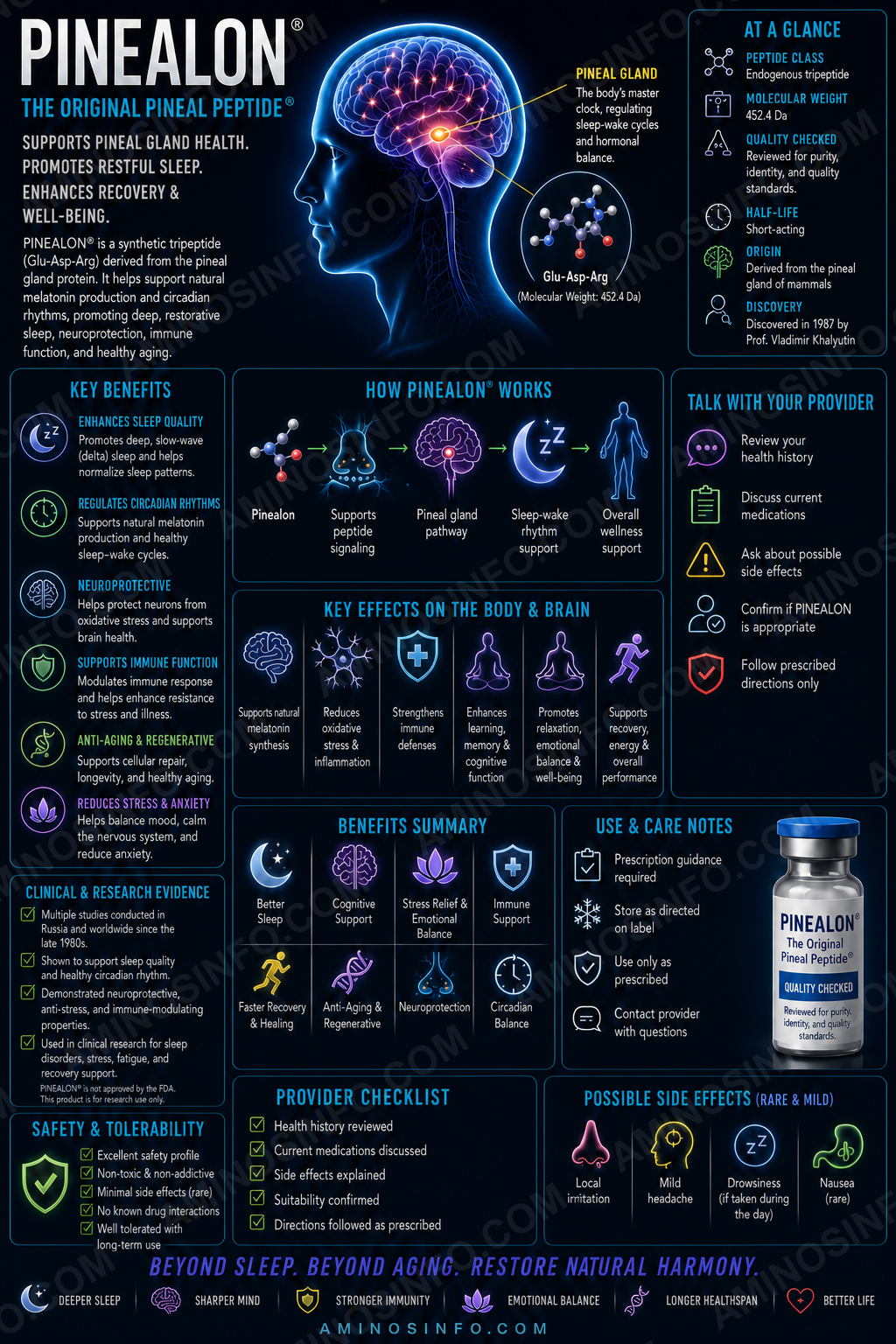
Pinealon, also called the EDR peptide, is a synthetic tripeptide composed of glutamic acid, aspartic acid, and arginine. It was developed within the Khavinson peptide-bioregulator research program and is commonly described as a neuroregulatory or brain-associated short peptide.
Researchers investigate Pinealon in relation to:
- Oxidative-stress resistance
- Hypoxia and ischemia tolerance
- Neuronal survival
- Learning and memory
- Dendritic-spine preservation
- Gene-expression regulation
- Age-related cognitive decline
- Experimental Alzheimer’s and Huntington’s disease biology
Glu-Asp-Arg
EDR
3 amino acids
C₁₅H₂₆N₆O₈
Approximately 418.40 g/mol
No
🧬 Molecular Structure
🧪 Amino-acid sequence
L-Glutamyl-L-Aspartyl-L-Arginine
Glu-Asp-Arg
EDR
Terminal chemistry
The standard reference compound is generally represented with a free N-terminal amino group and a free C-terminal arginine carboxyl group:
H-Glu-Asp-Arg-OH
Structural characteristics
- Three standard L-amino acids
- Linear tripeptide
- No cysteine residues
- No disulfide bonds
- Two acidic residues and one basic residue
- Highly polar and water-soluble free peptide
⚛️ Molecular Weight and 🧫 Formula
| Molecular formula | C15H26N6O8 |
|---|---|
| Average molecular weight | Approximately 418.40 g/mol |
| Monoisotopic mass | Approximately 418.1802 Da |
| Common CAS number | 175175-23-2 |
| PubChem CID | 10273502 |
| Disulfide bonds | None |
Salt and hydrate forms
Acetate, trifluoroacetate, sodium, or hydrated material can have a different apparent formula weight. A COA should state whether the reported quantity refers to free peptide, total salt mass, or lyophilized vial mass.
📅 Discovery Timeline and Research History
Late 20th century: Tissue-specific peptide research
Russian gerontology and military-medical laboratories investigated short peptides isolated from organ-derived peptide complexes.
Cortexin fractionation
EDR was described as a short peptide associated with neuroprotective fractions derived from the broader Cortexin cerebral-cortex peptide complex.
2000s: Hypoxia and aging studies
Pinealon was evaluated in hypobaric hypoxia, prenatal hypoxia, carotid-occlusion, aging, and cognitive models.
2008: Antihypoxic properties published
A study reported that Pinealon produced the strongest antihypoxic effect among several tested short peptides.
2011: Oxidative-stress and cell-viability study
Pinealon reduced reactive oxygen species and necrotic cell death in cerebellar granule, PC12, and immune-cell systems.
2012: Prenatal hyperhomocysteinemia study
Maternal treatment was associated with improved offspring cognition and increased cerebellar-neuron resistance to oxidative stress.
2014: Serotonin-expression research
Short peptides including EDR were reported to influence serotonin expression in cultured brain-cortex cells.
2015: Small human aging study
A 32-person regional study evaluated Pinealon and Vesugen using biological-age and metabolic markers, with mixed findings including possible pro-oxidant signals and reduced CD34-positive cell markers.
2020–2022: Gene-regulation and Alzheimer’s-mechanism publications
Studies and reviews proposed interactions with DNA, histones, gene promoters, and proteins related to synaptic plasticity and neurodegeneration.
Current status
Pinealon remains investigational and lacks large, independently replicated, blinded human trials or FDA approval.
Cortexin and Peptide-Bioregulator Origins
Cortexin relationship
Cortexin is a heterogeneous animal-derived cerebral-cortex peptide mixture. Pinealon is a single synthetic tripeptide proposed to reproduce part of the biological activity associated with that mixture.
Not a pineal-gland peptide
Despite its name, Pinealon is usually discussed as a brain/cortex neuroregulatory peptide rather than a defined natural pineal hormone.
Khavinson bioregulator framework
The broader theory proposes that ultrashort peptides can influence tissue-specific gene expression, protein synthesis, and cellular resilience.
Origin versus proof
Being derived conceptually from an active tissue fraction does not prove that the isolated tripeptide reproduces all Cortexin effects or acts selectively in the brain.
🧠 Proposed Mechanism of Action
1. Cellular uptake
Ultrashort peptides can enter cells through peptide transporters, endocytosis, or concentration-dependent membrane processes, but Pinealon’s dominant human transport route is not established.
2. Antioxidant effects
Pinealon reduced reactive oxygen species in several cell types exposed to receptor-dependent and direct oxidant stress.
3. Cell-survival effects
Reduced necrotic death and increased viability were reported in neuronal and PC12 systems.
4. Gene-expression regulation
Research from the Khavinson group proposes that EDR can interact with DNA- or chromatin-related systems and alter transcription of genes involved in neuronal function.
5. Synaptic and neurotransmitter pathways
Studies report effects on dendritic spines, serotonin expression, NMDA-receptor-subunit genes, and proteins implicated in Alzheimer’s disease.
🎯 Target and Pathway Profile
| Target or pathway | Evidence status |
|---|---|
| Single receptor | None established. |
| Reactive oxygen species | Reduced in several in-vitro systems. |
| Apoptosis and necrosis | Reduced in selected cellular and animal models. |
| DNA and histones | Direct or sequence-specific interactions are proposed; clinical significance remains uncertain. |
| Serotonin expression | Increased in cultured brain-cortex cells in one research program. |
| NMDA receptor genes | Altered in diabetic-rat hippocampal research. |
| Dendritic-spine preservation | Reported in amyloid and mutant-protein neuronal models. |
| Validated human biomarker | None established. |
Gene Expression and Epigenetic Hypotheses
DNA interaction
Computational, biophysical, and cell studies propose that EDR may interact with selected DNA sequences or regulatory regions.
Histone interaction
Ultrashort peptides are hypothesized to bind histones or affect chromatin accessibility, thereby changing transcription.
Protein-synthesis regulation
Some studies report increased expression of neuronal proteins involved in differentiation, synaptic plasticity, antioxidant defense, and aging.
Alzheimer’s-related genes
Published work proposes effects on genes or proteins associated with amyloid processing, tau biology, autophagy, and neuronal survival.
Mechanistic caution
Changes in mRNA or protein expression do not prove direct gene targeting. Secondary stress responses, altered cell viability, or pathway cross-talk can produce similar observations.
Independent replication need
Many gene-regulation claims come from one connected research network and require broader independent confirmation.
Oxidative-Stress and Cell-Survival Research
Reactive oxygen species
Pinealon reduced ROS accumulation induced by hydrogen peroxide, ouabain, homocysteine, and other stressors.
Cell models
Effects were reported in rat cerebellar granule cells, PC12 pheochromocytoma cells, and neutrophils.
Necrotic death
Propidium-iodide assays suggested lower necrotic cell death under selected conditions.
Proliferative activity
Some experiments reported increased proliferation or viability, which may support repair but also requires long-term growth-safety evaluation.
Antioxidant versus pro-oxidant findings
A small human geroprotection study reported possible pro-oxidant chemiluminescence effects, showing that biological responses may depend on dose, tissue, or disease state.
Hypoxia and Ischemia Research
Hypobaric hypoxia
Pinealon showed antihypoxic activity in animal models simulating low-oxygen exposure.
Prenatal hypoxia
Researchers reported improved resistance of offspring neurons after peptide exposure in developmental hypoxia paradigms.
Carotid-artery occlusion
Short-peptide administration before experimental carotid occlusion was associated with behavioral and apoptosis-related changes in old rats.
Mechanistic possibilities
- Reduced oxidative stress
- Improved mitochondrial resilience
- Altered stress-response gene expression
- Reduced caspase activation
- Preservation of synaptic function
No stroke-treatment evidence
Pinealon has no established role in emergency stroke care and does not replace thrombolysis, thrombectomy, antiplatelet therapy, vascular prevention, or rehabilitation.
Learning, Memory, and Cognition Research
Aging models
Regional studies report improvements in memory, attention, emotional state, or cognitive performance in older animals and selected patient groups.
Prenatal stress models
Offspring exposed to maternal hyperhomocysteinemia showed improved learning after Pinealon treatment in one rat study.
Diabetes models
Pinealon preserved learning and altered hippocampal NMDA-receptor-subunit gene expression in diabetic rats.
Professional-driver study
A regional study evaluated peptide correction of neurotic or fatigue-related symptoms in truck drivers, but methodological details and international replication are limited.
No healthy-person nootropic evidence
No large, blinded, placebo-controlled human study establishes improved memory, focus, reaction time, executive function, or academic performance.
Alzheimer’s and Huntington’s-Model Research
Amyloid synaptotoxicity
EDR and related peptides were reported to reduce dendritic-spine loss in neuronal cultures exposed to Alzheimer’s-related pathology.
Huntington’s disease models
Dendritic-spine preservation was also reported in cultures containing mutant huntingtin-related pathology.
Protein-expression studies
Research proposes regulation of proteins involved in synaptic maintenance, autophagy, apoptosis, and neuronal differentiation.
5xFAD work
Related short peptides have been evaluated in transgenic Alzheimer’s mice, although not every result can be attributed specifically to Pinealon.
No human disease-modifying evidence
Pinealon has not been shown to improve clinical dementia, reduce amyloid PET, lower tau biomarkers, prevent brain atrophy, or delay institutionalization.
Prenatal Hyperhomocysteinemia Research
Experimental design
Pregnant rats received high dietary methionine to induce hyperhomocysteinemia and oxidative stress.
Offspring findings
Pinealon treatment was associated with improved cognitive performance and increased cerebellar-neuron resistance to oxidative injury.
Potential pathways
- Reduced ROS
- Improved neuronal survival
- Altered developmental gene expression
- Protection from homocysteine toxicity
Pregnancy caution
Animal developmental findings do not establish safety in human pregnancy. Pinealon should not be promoted as prenatal neuroprotection.
Diabetes-Related Hippocampal Research
Experimental diabetes
Diabetic rats can develop oxidative stress, hippocampal dysfunction, and impaired learning.
Learning retention
Pinealon was reported to preserve or improve learning performance in one experimental-diabetes study.
NMDA-receptor genes
Changes were reported in expression of selected NMDA receptor subunits in the hippocampus.
No diabetes therapy
Pinealon is not an approved treatment for hyperglycemia, insulin resistance, diabetic neuropathy, or cognitive impairment associated with diabetes.
Serotonin and Neurotransmitter Research
Serotonin expression
Short peptides including Pinealon were reported to stimulate serotonin expression in cultured cortical cells.
Not an SSRI
Pinealon has not been shown to directly inhibit the serotonin transporter or act like fluoxetine, sertraline, or other SSRIs.
Not a serotonin receptor agonist
No validated serotonin-receptor binding profile has been established.
Behavioral implications
Changes in serotonin-related proteins may influence mood or cognition, but cell-expression findings cannot predict clinical antidepressant effects.
Healthy-Aging and Clinical Claims
Organic brain syndrome and polymorbidity
A small regional study examined Pinealon and Vesugen in adults aged 41–83 with chronic polymorbidity and organic brain syndrome in remission.
Reported findings
Investigators reported changes in biological-age and CNS-function indicators.
Concerning findings
The same abstract described pro-oxidant activity and reduced circulating CD34-positive cells, complicating a uniformly beneficial interpretation.
Study limitations
- Only 32 participants
- Unclear randomization and blinding
- Multiple biomarkers
- No modern clinical-outcome endpoint
- Limited independent replication
No established longevity effect
There is no evidence that Pinealon extends human lifespan, prevents dementia, reverses biological age, or reduces all-cause mortality.
Evidence Limitations and Clinical Interpretation
Research-group concentration
Many foundational papers were authored by researchers affiliated with the same peptide-bioregulator program.
Preclinical dominance
Most evidence comes from cells and animals rather than controlled human trials.
Broad mechanistic claims
DNA binding, histone regulation, antioxidant effects, serotonin expression, dendritic-spine preservation, and antihypoxic activity may not all occur at the same exposure or in the same tissue.
Dose uncertainty
Human pharmacokinetics, oral bioavailability, tissue distribution, brain exposure, active metabolites, and optimal dose are not well defined.
Clinical-outcome gap
Changes in ROS, gene expression, dendritic spines, or biological-age markers do not establish improved survival, dementia prevention, or functional independence.
Publication and language bias
Regional and Russian-language literature may be difficult to verify fully and may underrepresent negative findings.
Safety and Regulatory Considerations
No established human safety profile
No FDA-approved label defines dosage, route, contraindications, interactions, pregnancy safety, or long-term adverse effects.
Potential neurological risks
- Headache
- Sleep or mood changes
- Agitation
- Altered neurotransmitter signaling
- Unknown seizure effects
Potential proliferative concerns
Reported increases in cell viability or proliferation require assessment for abnormal-cell survival and long-term cancer relevance.
Potential oxidative concerns
Although many studies report antioxidant activity, at least one small human study reported possible pro-oxidant signals.
Pregnancy and pediatric use
Animal developmental research does not establish human safety in pregnancy, infants, or children.
Drug interactions
Interactions with antidepressants, antipsychotics, antiseizure drugs, anticoagulants, diabetes medicines, or chemotherapy are unknown.
Regulatory status
Pinealon is not FDA approved.
🧪 Laboratory Testing Methods
| Method | Purpose | Important limitation |
|---|---|---|
| RP-HPLC / UPLC | Separates EDR from deletion products, free amino acids, and degradants. | Area purity does not prove sequence or net content. |
| LC-HRMS | Confirms intact mass and elemental composition. | Sequence isomers and epimers can share mass. |
| MS/MS sequencing | Confirms Glu-Asp-Arg residue order. | Requires validated fragmentation interpretation. |
| Edman degradation | Orthogonally confirms N-terminal sequence. | Less useful for low-level impurities. |
| Amino-acid analysis | Confirms E:D:R composition and supports content assignment. | Does not prove residue order. |
| Chiral amino-acid analysis | Confirms L-amino-acid configuration and detects epimers. | Hydrolysis can introduce artifacts. |
| Aspartimide and isoaspartate assay | Detects Asp-related synthesis and storage degradants. | Requires specialized LC-MS methods. |
| Pyroglutamate assay | Checks for N-terminal glutamate cyclization. | May require targeted mass analysis. |
| Net peptide-content assay | Measures actual EDR quantity. | Must correct for water, counterions, and residual solvents. |
| Residual-solvent and counterion testing | Quantifies TFA, acetate, acetonitrile, and other process residues. | Does not establish biological activity. |
| ROS-reduction assay | Measures one reported functional effect. | Highly dependent on cell type and stressor. |
| Cell-viability and necrosis assays | Evaluate protective activity. | No internationally validated Pinealon potency assay exists. |
| Gene-expression panel | Measures selected neuronal and stress-response transcripts. | Expression changes do not prove direct DNA targeting. |
| DNA/histone-binding assay | Tests proposed epigenetic interactions. | In-vitro binding may not occur at physiological exposure. |
| Plasma and protease stability | Measures degradation and metabolites. | Animal matrices do not fully predict humans. |
| Brain/plasma pharmacokinetics | Measures systemic and CNS exposure. | Human data remain inadequate. |
| Microbial limits, sterility, and endotoxin | Evaluate route-specific microbiological quality. | Requirements depend on final dosage form. |
| Stability-indicating assay | Tracks hydrolysis, cyclization, epimerization, oxidation, and potency loss. | Requires qualified reference standards. |
📄 How to Interpret a Pinealon COA
- Verify the exact sequence: Glu-Asp-Arg, or EDR.
- Confirm the free-peptide formula and mass: C₁₅H₂₆N₆O₈ and approximately 418.40 g/mol.
- Confirm terminal chemistry: H-Glu-Asp-Arg-OH unless a modified analogue is explicitly intended.
- Use MS/MS or Edman sequencing: HPLC and intact mass alone cannot prove residue order.
- Confirm L-stereochemistry: D-amino-acid epimers may have the same mass but different biology.
- Review Asp-related degradation and N-terminal pyroglutamate formation.
- Measure net peptide content: “99% purity” is not the labeled number of milligrams.
- Report counterions, water, and residual solvents separately.
- Require a validated functional assay: ROS, viability, or gene-expression assays can support potency, but none proves clinical efficacy.
- Do not infer medical benefit: A COA cannot prove memory improvement, neuroprotection, Alzheimer’s prevention, lifespan extension, or human safety.
📊 Pinealon vs Cortagen vs Cortexin vs Semax
| Feature | Pinealon | Cortagen | Cortexin | Semax |
|---|---|---|---|---|
| Structure | EDR tripeptide | AEDP tetrapeptide | Animal-derived peptide mixture | Defined synthetic heptapeptide analogue |
| Main focus | Neuroprotection and gene regulation | Cortical resilience and nerve repair | Broad neuroprotection | Neurotrophic and cognitive signaling |
| Single sequence? | Yes | Yes | No | Yes |
| FDA approved? | No | No | No | No |
Pinealon vs Epitalon vs Vilon vs Vesugen
| Peptide | Sequence | Main research focus |
|---|---|---|
| Pinealon | EDR | Brain, oxidative stress, cognition |
| Epitalon | AEDG | Healthy aging and circadian biology |
| Vilon | KE | Immune and chromatin signaling |
| Vesugen | KED | Vascular and endothelial signaling |
Pinealon vs P021 vs FGL vs DNSP-11
| Compound | Main proposed mechanism | Main research theme |
|---|---|---|
| Pinealon | Gene regulation and antioxidant signaling | Neuronal resilience and cognition |
| P021 | LIF/BDNF/GSK3β-related signaling | Neurogenesis, tau, memory |
| FGL | NCAM–FGFR1 signaling | Synaptic plasticity |
| DNSP-11 | ERK and mitochondrial protection | Dopaminergic-neuron support |
Pinealon vs Evidence-Based Cognitive Care
| Approach | Established role | Difference from Pinealon |
|---|---|---|
| Exercise, sleep, hearing correction, vascular control | Supports cognitive health | Human evidence and known safety |
| Cholinesterase inhibitors | Symptomatic treatment in selected dementias | Approved human medicines |
| Anti-amyloid antibodies | Selected early Alzheimer’s disease | Human biomarker and outcome evidence |
| Pinealon | Experimental ultrashort peptide | No established human efficacy or long-term safety |
🔗 Related Peptides and Pathways
- Cortexin: Animal-derived cortex peptide complex associated with Pinealon’s discovery.
- Cortagen: AEDP cortex-related tetrapeptide.
- Epitalon: AEDG aging-related tetrapeptide.
- KED peptide: Neurovascular tripeptide studied alongside EDR.
- NMDA receptors: Hippocampal gene-expression target in diabetic-rat research.
- Serotonin: Expression altered in cortical-cell studies.
- Reactive oxygen species: Reduced in several cell models.
- DNA and histones: Proposed gene-regulatory interaction partners.
🖼️ Original Diagram Specifications
Diagram 1: Pinealon molecular sequence
Show Glu-Asp-Arg with free N- and C-termini, acidic side chains, arginine guanidinium group, formula, and molecular weight.
Diagram 2: Cortexin-to-Pinealon development
Show the heterogeneous Cortexin mixture, fractionation, identification of EDR, and synthetic Pinealon.
Diagram 3: Proposed gene-regulation pathway
Show EDR entering a cell, proposed DNA/histone interaction, transcriptional changes, protein synthesis, and neuronal outcomes, with the pathway labeled “hypothesis under investigation.”
Diagram 4: Oxidative-stress protection
Show hydrogen peroxide or homocysteine increasing ROS, mitochondrial injury, necrosis, and Pinealon-associated reduction in oxidative damage.
Diagram 5: Hypoxia research
Show low oxygen, mitochondrial stress, caspase activation, behavioral deficits, and the preclinical Pinealon findings.
Diagram 6: Evidence ladder
Show chemistry, cell studies, rodent studies, small regional human studies, independent phase I, phase II, phase III, and approval.
Diagram 7: COA workflow
Show exact sequence, stereochemistry, HRMS, MS/MS, Asp degradation, pyroglutamate, net content, counterions, functional assay, microbiology, and stability.
❓ Frequently Asked Questions
Is Pinealon a peptide?
Yes. Pinealon is a synthetic tripeptide composed of glutamic acid, aspartic acid, and arginine.
What is its exact sequence?
Glu-Asp-Arg, abbreviated EDR.
What is its molecular formula?
C₁₅H₂₆N₆O₈ for the free peptide.
What is its molecular weight?
Approximately 418.40 g/mol.
What is its CAS number?
175175-23-2 is commonly used.
Is Pinealon derived from the pineal gland?
It is more commonly described as a cortex-associated peptide developed from Cortexin-related research, despite its name.
Is Pinealon the same as Cortexin?
No. Cortexin is a complex animal-derived mixture; Pinealon is one defined synthetic tripeptide.
Is Pinealon the same as Cortagen?
No. Cortagen is Ala-Glu-Asp-Pro, whereas Pinealon is Glu-Asp-Arg.
What is Pinealon studied for?
Oxidative stress, hypoxia, cognition, neuronal survival, dendritic spines, gene expression, and healthy-aging models.
Does Pinealon improve memory?
Several animal and small regional studies report cognitive effects, but robust human evidence is lacking.
Does Pinealon treat Alzheimer’s disease?
No human treatment benefit has been established.
Does Pinealon increase serotonin?
One research program reported increased serotonin expression in cultured cortical cells. It is not an established serotonin drug.
Does Pinealon extend lifespan?
No human lifespan extension has been demonstrated.
Is Pinealon FDA approved?
No.
Could Pinealon have pro-oxidant effects?
Most preclinical work reports antioxidant effects, but one small human study described possible pro-oxidant findings.
Does 99% HPLC purity prove authentic Pinealon?
No. Sequence, stereochemistry, mass, degradation products, net content, and functional activity require separate confirmation.
Final Thoughts
Pinealon is a chemically defined three-amino-acid peptide with the sequence Glu-Asp-Arg. Its free-peptide formula is C₁₅H₂₆N₆O₈ and its average molecular weight is approximately 418.40 g/mol.
Preclinical research reports reduced oxidative stress, improved cell survival, antihypoxic activity, altered gene expression, dendritic-spine preservation, serotonin-related changes, and improved cognition in selected animal models. However, most findings come from a concentrated regional research program, and high-quality independent human evidence remains limited.
Legitimate Pinealon material should be tested for exact EDR sequence, L-stereochemistry, molecular mass, Asp-related degradation, pyroglutamate formation, residual solvents, counterions, water, net peptide quantity, relevant functional activity, route-specific microbiological quality, and stability. Analytical purity cannot establish memory improvement, neuroprotection, dementia prevention, longevity, or human safety.
📚 References
- Arutjunyan A, et al. Pinealon protects rat offspring from prenatal hyperhomocysteinemia. International Journal of Clinical and Experimental Medicine. 2012.
- Khavinson V, et al. EDR Peptide: Possible Mechanism of Gene Expression and Protein Synthesis Regulation Involved in the Pathogenesis of Alzheimer’s Disease. Molecules. 2020.
- PubChem. Glu-Asp-Arg compound record.
- Kozina LS, et al. Investigation of antihypoxic properties of short peptides. Advances in Gerontology. 2008.
- Khavinson V, et al. Pinealon increases cell viability by suppression of free radical levels and activating proliferative processes. Rejuvenation Research. 2011.
- Khavinson V, et al. Neuroprotective Effects of Tripeptides—Epigenetic Regulators of Gene Expression. Pharmaceuticals. 2021.
- Ilina A, et al. Neuroepigenetic Mechanisms of Action of Ultrashort Peptides in Alzheimer’s Disease. International Journal of Molecular Sciences. 2022.
- Meshchaninov VN, et al. Effect of synthetic peptides on aging of patients with chronic polymorbidity and organic brain syndrome. Advances in Gerontology. 2015.
- Khavinson VK, et al. Short peptides stimulate serotonin expression in cells of brain cortex. Bulletin of Experimental Biology and Medicine. 2014.
- Khavinson VK, et al. Effect of Pinealon on learning and expression of NMDA receptor subunit genes in the hippocampus of rats with experimental diabetes. Neurochemical Journal. 2020.
- Khavinson VK, et al. Effects of short peptides before carotid artery occlusion on behavior and caspase-3 activity in old rats. Advances in Gerontology. 2011.
- Khavinson VK, et al. Peptide correction of neurotic disorders among professional truck drivers. Advances in Gerontology. 2012.
- Khavinson VK. Peptide regulation of aging. Human Physiology.
- Khavinson VK, et al. Peptide bioregulators: the new class of geroprotectors. Advances in Gerontology.
- Khavinson VK, et al. Tissue-specific effects of short peptides and regulation of gene expression. Neuroendocrinology Letters.
- Khavinson VK, et al. Short peptides and epigenetic regulation of aging. Advances in Gerontology.
- Khavinson V, et al. Peptides and C-terminal fragments in neuroprotection. Journal of Amino Acids.
- Fedoreyeva LI, et al. Short peptides and DNA interaction. Biochemistry Moscow.
- Ashapkin VV, et al. Epigenetic mechanisms of geroprotective peptides. Advances in Gerontology.
- Linkova NS, et al. Short peptides regulate gene expression in nervous tissue. Bulletin of Experimental Biology and Medicine.
- Polyakova VO, et al. Effects of EDR and KED peptides on neuronal proteins and synaptic plasticity. Advances in Gerontology.
- Safarova ER, et al. EDR peptide and dendritic-spine preservation in amyloid synaptotoxicity. Experimental Gerontology.
- Khavinson V, et al. Peptide regulation of neuronal differentiation and aging. Cellular and Molecular Neurobiology.
- Mattson MP. Pathways towards and away from Alzheimer’s disease. Nature.
- Long JM, Holtzman DM. Alzheimer disease: an update on pathobiology and treatment strategies. Cell.
- Masters CL, et al. Alzheimer’s disease. Nature Reviews Disease Primers.
- Haass C, Selkoe DJ. Soluble protein oligomers in neurodegeneration. Nature Reviews Molecular Cell Biology.
- Ross CA, Tabrizi SJ. Huntington’s disease: from molecular pathogenesis to clinical treatment. Lancet Neurology.
- Bates GP, et al. Huntington disease. Nature Reviews Disease Primers.
- Lau A, Tymianski M. Glutamate receptors, neurotoxicity and neurodegeneration. Pflügers Archiv.
- Hardingham GE, Bading H. Synaptic versus extrasynaptic NMDA receptor signalling. Nature Reviews Neuroscience.
- Traynelis SF, et al. Glutamate receptor ion channels: structure, regulation, and function. Pharmacological Reviews.
- Murphy MP. How mitochondria produce reactive oxygen species. Biochemical Journal.
- Sies H, Jones DP. Reactive oxygen species as pleiotropic physiological signalling agents. Nature Reviews Molecular Cell Biology.
- Radi E, et al. Apoptosis and oxidative stress in neurodegenerative diseases. Journal of Neurochemistry.
- Green DR, Kroemer G. The pathophysiology of mitochondrial cell death. Science.
- Elmore S. Apoptosis: a review of programmed cell death. Toxicologic Pathology.
- Mattson MP, Shea TB. Folate and homocysteine metabolism in neural plasticity and neurodegenerative disorders. Trends in Neurosciences.
- Smith AD, Refsum H. Homocysteine, B vitamins, and cognitive impairment. Annual Review of Nutrition.
- Moretti R, Caruso P. The controversial role of homocysteine in neurology. International Journal of Molecular Sciences.
- Dirnagl U, Iadecola C, Moskowitz MA. Pathobiology of ischemic stroke. Trends in Neurosciences.
- Lo EH. A new penumbra: transitioning from injury into repair after stroke. Nature Medicine.
- van der Flier WM, et al. Vascular cognitive impairment. Nature Reviews Disease Primers.
- Biessels GJ, Despa F. Cognitive decline and dementia in diabetes mellitus. Nature Reviews Endocrinology.
- Biessels GJ, Reagan LP. Hippocampal insulin resistance and cognitive dysfunction. Nature Reviews Neuroscience.
- Haroon E, et al. Concepts and neurobiology of fatigue and stress-related cognitive dysfunction. Neuropsychopharmacology.
- Berger M, Gray JA, Roth BL. The expanded biology of serotonin. Annual Review of Medicine.
- Müller CP, Cunningham KA. Handbook of the Behavioral Neurobiology of Serotonin.
- Fosgerau K, Hoffmann T. Peptide therapeutics: current status and future directions. Drug Discovery Today.
- Lau JL, Dunn MK. Therapeutic peptides: historical perspectives and current development trends. Bioorganic & Medicinal Chemistry.
- Craik DJ, Fairlie DP, Liras S, Price D. The future of peptide-based drugs. Chemical Biology & Drug Design.
- International Council for Harmonisation. ICH Q2(R2): Validation of Analytical Procedures.
- International Council for Harmonisation. ICH Q3A and Q3B: Impurities in New Drug Substances and Products.
- International Council for Harmonisation. ICH Q3C: Residual Solvents.
- International Council for Harmonisation. ICH Q1A(R2): Stability Testing of New Drug Substances and Products.
- International Council for Harmonisation. ICH M10: Bioanalytical Method Validation.
- United States Pharmacopeia General Chapter <621>: Chromatography.
- United States Pharmacopeia General Chapter <71>: Sterility Tests.
- United States Pharmacopeia General Chapter <85>: Bacterial Endotoxins Test.
- United States Pharmacopeia General Chapters <232> and <233>: Elemental Impurities.
Identity, sequence, chemistry, Cortexin origins, oxidative stress, hypoxia, cognition, gene regulation, Alzheimer’s models, serotonin, aging, safety, and analytical evidence were reviewed in July 2026. Pinealon remains an unapproved investigational peptide.