Dihexa (PNB-0408): What It Is, How It Works, Benefits, and Research Overview
A corrected, evidence-graded review of Dihexa, including its angiotensin-IV-derived chemical structure, molecular properties, cognition and neuroprotection studies, disputed HGF/c-Met mechanism, the 2025 retraction of the foundational synaptogenesis paper, Alzheimer’s-model research, cancer concerns, safety, analytical testing, and COA interpretation.
What Is Dihexa?
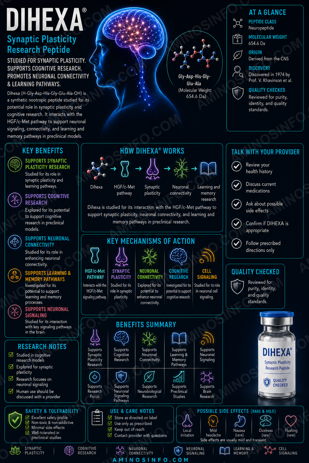
Dihexa, also known as PNB-0408, is a synthetic, lipid-modified angiotensin-IV-derived peptidomimetic developed for experimental cognition and neurorepair research.
It is commonly described as:
N-hexanoyl-Tyr-Ile-(6-aminohexanoic amide)
Dihexa is not a conventional peptide composed only of standard amino acids. It contains:
- An N-terminal hexanoyl lipid chain
- L-tyrosine
- L-isoleucine
- 6-aminohexanoic acid
- A terminal carboxamide
PNB-0408
Angiotensin-IV-derived peptidomimetic
C₂₇H₄₄N₄O₅
Approximately 504.67 g/mol
1401708-83-5
No
🧬 Molecular Structure
🧪 Structural description
Hexanoyl-L-Tyr-L-Ile-6-aminohexanamide
IUPAC name
6-[(2S,3S)-2-[(2S)-2-hexanamido-3-(4-hydroxyphenyl)propanamido]-3-methylpentanamido]hexanamide
SMILES
CCCCCC(=O)N[C@@H](Cc1ccc(O)cc1)C(=O)N[C@@H]([C@@H](C)CC)C(=O)NCCCCCC(N)=O
⚛️ Molecular Weight and 🧫 Formula
| Molecular formula | C27H44N4O5 |
|---|---|
| Average molecular weight | Approximately 504.67 g/mol |
| Monoisotopic mass | Approximately 504.3312 Da |
| Common CAS number | 1401708-83-5 |
| Stereochemistry | L-tyrosine and L-isoleucine configuration must be preserved |
| Disulfide bonds | None |
| Ionizable features | Phenolic hydroxyl and terminal amide; no free terminal carboxylate |
Why the hexanoyl group matters
The lipid chain increases hydrophobicity and was intended to improve metabolic stability, oral activity, and access to the central nervous system compared with unmodified angiotensin IV.
Why 6-aminohexanamide matters
The flexible aminohexanoic-amide segment replaces the C-terminal portion of angiotensin IV and contributes to resistance against peptidases.
📅 Discovery Timeline and Research History
1980s–1990s: Angiotensin IV cognition research
Angiotensin IV was found to influence learning, memory, cerebral blood flow, and hippocampal function in animal studies.
2000s: Stable AngIV analogues developed
Researchers created metabolically stable analogues such as Nle¹-AngIV and C-terminally truncated compounds.
2011: Truncated analogues and memory
C-terminally modified angiotensin-IV analogues improved spatial memory and synaptogenesis-related measures in rodent studies.
2014: Dihexa HGF/c-Met paper published
A paper reported HGF binding, c-Met dependence, dendritic-spine growth, and synaptogenesis.
2015: Reviews and hair-cell protection
Dihexa was reviewed as a potential orally active neurotrophic compound, and a zebrafish study reported protection against aminoglycoside-induced lateral-line hair-cell injury.
2021: APP/PS1 mouse study
A study reported improved spatial learning, neuronal markers, inflammation, apoptosis, and PI3K/AKT signaling in an Alzheimer’s mouse model.
2024: Mitochondrial-toxin research
An angiotensin-IV analogue identified as Dihexa was evaluated in 3-nitropropionic-acid-related cellular injury.
2025: Foundational mechanism paper retracted
The 2014 HGF/c-Met synaptogenesis paper received a formal retraction notice. This materially lowers confidence in the most frequently cited mechanism.
Current status
No established Dihexa phase 1–3 human clinical program or FDA-approved Dihexa product exists.
Angiotensin IV Origins and Analogue Design
Angiotensin IV
Angiotensin IV is the hexapeptide Val-Tyr-Ile-His-Pro-Phe, generated within the renin–angiotensin system.
AT4 receptor concept
Early research proposed a distinct “AT4 receptor” responsible for cognitive effects. Insulin-regulated aminopeptidase was later identified as a high-affinity binding protein for many AngIV analogues.
Nle¹-AngIV
Substitution and truncation studies produced more stable analogues with procognitive activity.
Dihexa design
Dihexa retains the Tyr-Ile core while adding a hexanoyl group and aminohexanamide tail to increase stability and lipophilicity.
Mechanistic uncertainty
AngIV analogues may influence IRAP, HGF/c-Met, cerebral blood flow, glucose transport, dopamine, and other systems. Dihexa’s definitive molecular target remains unresolved after the retraction.
🧠 Proposed Mechanism of Action
Historical HGF/c-Met claim RETRACTED FOUNDATION
The 2014 study reported that Dihexa bound HGF, potentiated subthreshold HGF, activated c-Met, increased dendritic spines and synapses, and lost activity when c-Met was inhibited.
Why the retraction matters
Retraction means the paper should not be relied upon as valid evidence. Reviews and vendor summaries that repeat its conclusions without noting the retraction are outdated.
What remains independently observed
- Cognitive improvement in selected rodent models
- Changes in inflammatory and apoptotic markers in APP/PS1 mice
- PI3K/AKT-associated effects in one mouse study
- Hair-cell protection in zebrafish
- Neuroprotection in selected toxin models
What remains unresolved
- Direct molecular target
- Whether HGF binding occurs reliably
- Whether c-Met activation is necessary
- Whether Dihexa directly induces human synaptogenesis
- Whether effects involve IRAP or other AngIV pathways
🎯 Target and Pathway Profile
| Target or pathway | Evidence status |
|---|---|
| HGF | Direct binding was reported in a paper retracted in 2025. |
| c-Met / MET | Dependency was reported in the retracted paper; mechanism is not secure. |
| PI3K/AKT | Supported by a 2021 APP/PS1 mouse study using pathway inhibition. |
| IRAP / LNPEP | Relevant to AngIV biology, but direct Dihexa pharmacology remains incompletely defined. |
| Synaptophysin | Increased in APP/PS1 mouse tissue in one study. |
| Inflammatory cytokines | Reduced in selected Alzheimer’s-model research. |
| Apoptosis markers | Reduced in selected cellular and mouse studies. |
| Validated human receptor | None established. |
HGF/c-Met Biology and Cancer Relevance
HGF and MET
Hepatocyte growth factor activates the MET receptor tyrosine kinase, regulating growth, motility, survival, tissue repair, angiogenesis, and development.
Neural functions
HGF/MET signaling participates in neurite growth, neuronal survival, synaptic development, and plasticity.
Oncogenic functions
MET is a proto-oncogene. Excessive HGF/MET signaling can promote tumor-cell proliferation, invasion, metastasis, angiogenesis, and resistance to therapy.
Mechanism uncertainty does not remove risk
Even though the direct Dihexa–HGF/c-Met evidence was retracted, any compound claimed to potentiate this pathway requires rigorous cancer and proliferative-safety testing.
No adequate carcinogenicity evidence
No long-term animal carcinogenicity program or human oncology-safety dataset has established that chronic Dihexa exposure is safe.
Learning and Cognition Research
AngIV analogue literature
Angiotensin IV and multiple analogues improved memory in normal and cognitively impaired rodents across spatial, passive-avoidance, and object-recognition paradigms.
Dihexa-specific evidence
Dihexa has been reported to improve performance in selected rodent cognitive models, but the direct evidence base is much smaller than marketing descriptions suggest.
Memory consolidation and retrieval
Reviews describe effects on both memory formation and recall, but many statements trace back to the retracted mechanism paper or connected laboratory work.
No healthy-person nootropic evidence
No controlled human study establishes improvement in memory, focus, IQ, executive function, or learning.
Behavioral-model limitations
Rodent maze performance can be affected by locomotion, anxiety, motivation, vision, stress, and sensory function—not cognition alone.
Alzheimer’s-Model Research
APP/PS1 mouse study
A 2021 study reported that Dihexa improved Morris-water-maze performance in APP/PS1 mice.
Neuronal and synaptic markers
The study reported increased neuronal-cell staining and synaptophysin expression.
Inflammation and apoptosis
Dihexa reduced selected inflammatory cytokines and apoptotic markers.
PI3K/AKT involvement
Wortmannin, a PI3K inhibitor, reversed several reported effects, supporting PI3K/AKT involvement in that model.
Angiotensin-IV levels
Tissue AngIV concentrations reportedly increased after Dihexa treatment, suggesting metabolism or pathway interactions that remain unclear.
No human Alzheimer’s evidence
Dihexa has not been shown to improve clinical cognition, daily function, amyloid PET, tau biomarkers, brain atrophy, or disease progression in humans.
Neuroprotection and Mitochondrial Research
3-Nitropropionic acid
Recent research examined Dihexa in a mitochondrial toxin model involving complex-II inhibition and oxidative stress.
Cell-survival pathways
Reported effects include reduced oxidative injury, inflammation, and apoptosis in selected experimental systems.
Neuroprotection versus neurotrophy
Preventing acute cell death is not equivalent to rebuilding lost neural circuits or reversing neurodegenerative disease.
No validated therapeutic window
The concentration required for neuroprotection relative to proliferative, hepatic, cardiovascular, or off-target effects is unknown.
Hair-Cell and Ototoxicity Research
Zebrafish lateral-line model
Dihexa protected lateral-line sensory hair cells against aminoglycoside exposure in zebrafish larvae.
HGF-mimetic interpretation
The study interpreted Dihexa as an HGF mimetic, based partly on the mechanism literature that is now weakened by retraction.
Model limitations
Zebrafish lateral-line cells are useful for ototoxicity screening but do not fully reproduce the human cochlea.
No hearing-loss treatment evidence
Dihexa has not been shown to prevent or reverse aminoglycoside hearing loss, tinnitus, or sensorineural hearing loss in humans.
PI3K/AKT and Inflammatory Pathways
PI3K/AKT
This pathway regulates survival, metabolism, protein synthesis, growth, and inflammation.
APP/PS1 evidence
Dihexa increased PI3K/AKT-related signaling in one mouse study, and pathway inhibition reduced reported benefits.
Inflammatory markers
Changes were reported in cytokines and glial-associated inflammatory responses.
Cancer overlap
PI3K/AKT is also frequently dysregulated in cancer. Chronic activation requires careful safety evaluation.
Pathway activation is not target identification
Observing PI3K/AKT changes does not reveal which receptor or direct molecular interaction initiated the effect.
Oral Activity and Brain-Penetration Claims
Design intent
The hexanoyl group and noncanonical tail were intended to improve metabolic stability and lipophilicity.
Oral activity in animals
Reviews and preclinical reports describe oral activity in rodent studies.
Blood–brain barrier claim
Dihexa is widely described as brain penetrant, but detailed peer-reviewed human pharmacokinetic and brain-exposure data are unavailable.
No human bioavailability data
Absorption, food effects, protein binding, metabolism, half-life, clearance, active metabolites, and CNS exposure are unknown in humans.
Topical and intranasal claims
Commercial use claims for topical or intranasal administration lack validated human pharmacology.
Cancer and Growth-Signaling Concerns
MET is a proto-oncogene
MET activation supports invasive growth and is a recognized driver or resistance pathway in multiple cancers.
HGF, PI3K, and AKT overlap
Pathways historically associated with Dihexa are also central to cancer-cell survival, proliferation, angiogenesis, and metastasis.
No proof of cancer safety
Statements that Dihexa cannot cause cancer because “multiple mutations are required” are not an adequate safety argument.
Potential high-risk populations
Particular caution is warranted conceptually in people with active cancer, prior cancer, premalignant lesions, unexplained masses, or ongoing chemotherapy.
Long-term exposure unknown
Neurotrophic compounds may require chronic use, making carcinogenicity, tumor-promotion, and abnormal-synapse research especially important.
Evidence Limitations and Clinical Interpretation
Foundational paper retracted
The strongest mechanistic and synaptogenesis claims relied on a paper that was retracted in 2025.
No human clinical trials
No validated dose-ranging, pharmacokinetic, safety, or efficacy study in humans has been established.
Small evidence base
Direct Dihexa studies are relatively few, and many reviews repeat the same foundational claims.
Connected authorship
Much of the early Dihexa research came from a connected group involved in developing and commercializing the compound.
Animal-model limitations
APP/PS1 mice, toxin models, and zebrafish hair cells do not reproduce the full complexity of human neurodegenerative disease.
Unknown target
Without a secure molecular target, potency assays, off-target screening, biomarker selection, and rational clinical dosing are difficult.
Safety and Regulatory Considerations
No established human safety profile
No approved label defines dose, route, contraindications, interactions, pregnancy safety, or chronic adverse effects.
Potential neurological risks
- Headache, agitation, insomnia, or mood changes
- Maladaptive synaptic growth
- Seizure or excitotoxicity risk
- Abnormal sensory processing
- Psychiatric effects
Potential proliferative risks
- Tumor promotion
- Angiogenesis
- Fibrosis
- Abnormal tissue growth
- Interaction with cancer therapy
Potential systemic risks
- Liver toxicity
- Cardiovascular effects
- Renal effects
- Immune reactions
- Unknown drug interactions
Product-quality risk
Unapproved material may contain incorrect stereochemistry, deletion products, residual protecting groups, oxidation products, hydrolysis products, endotoxin, residual solvents, or inaccurate content.
Regulatory status
Dihexa is not FDA approved.
🧪 Laboratory Testing Methods
| Method | Purpose | Important limitation |
|---|---|---|
| RP-HPLC / UPLC | Separates Dihexa from synthetic impurities and degradants. | Area purity does not prove identity or stereochemistry. |
| LC-HRMS | Confirms intact mass and elemental composition. | Epimers may share the same mass. |
| MS/MS fragmentation | Confirms sequence-like connectivity and terminal groups. | Noncanonical linker fragmentation requires interpretation. |
| 1D and 2D NMR | Confirms chemical structure, hexanoyl group, aminohexanamide tail, and connectivity. | Requires a qualified reference standard. |
| Chiral HPLC or Marfey analysis | Confirms L-Tyr and L-Ile stereochemistry. | Hydrolysis-based methods can introduce artifacts. |
| Residual protecting-group assay | Detects synthesis-related aromatic and carbamate impurities. | Requires process-specific knowledge. |
| Water and residual-solvent testing | Measures nonactive mass and solvent contamination. | Does not establish biological activity. |
| Net content by quantitative NMR or validated assay | Measures actual Dihexa content. | HPLC purity alone cannot provide net content. |
| HGF-binding assay | Historically used to evaluate the proposed mechanism. | Must be revalidated independently because foundational evidence was retracted. |
| c-Met phosphorylation assay | Tests pathway activation in a cell system. | Activation may be indirect and cell-line dependent. |
| PI3K/AKT assay | Measures downstream pathway effects. | Not specific to Dihexa or a particular receptor. |
| Broad kinase and receptor panel | Evaluates off-target pharmacology. | Requires clinically relevant concentrations. |
| Genotoxicity and proliferation assays | Assess DNA damage and growth stimulation. | Short in-vitro assays cannot replace long-term carcinogenicity studies. |
| Plasma and microsomal stability | Measures metabolism and active metabolites. | Animal matrices do not fully predict humans. |
| Brain/plasma pharmacokinetics | Measures systemic and CNS exposure. | No established human data. |
| Microbial limits, sterility, and endotoxin | Evaluate route-specific microbiological quality. | Requirements depend on final dosage form. |
| Stability-indicating assay | Tracks hydrolysis, oxidation, epimerization, aggregation, and potency loss. | Requires validated reference materials. |
📄 How to Interpret a Dihexa COA
- Verify the exact chemical structure: Hexanoyl-L-Tyr-L-Ile-6-aminohexanamide.
- Confirm formula and molecular weight: C₂₇H₄₄N₄O₅ and approximately 504.67 g/mol.
- Confirm stereochemistry: L-Tyr and L-Ile must be verified.
- Require NMR and LC-HRMS: HPLC alone is inadequate for a modified peptidomimetic.
- Review residual protecting groups, deletion products, unacylated material, hydrolysis products, epimers, water, and residual solvents.
- Measure net content: “99% purity” is not the labeled number of milligrams.
- Require independent functional testing: Any HGF/c-Met potency claim should acknowledge the 2025 retraction and use independently validated methods.
- Review proliferation and genotoxicity data: A routine peptide COA does not address cancer risk.
- Match microbiological testing to route: Finished intranasal or injectable forms require route-specific controls.
- Do not infer efficacy: A COA cannot prove memory improvement, synaptogenesis, brain penetration, Alzheimer’s treatment, or safety.
📊 Dihexa vs Angiotensin IV vs Nle¹-AngIV
| Feature | Dihexa | Angiotensin IV | Nle¹-AngIV |
|---|---|---|---|
| Structure | Lipid-modified peptidomimetic | Six-amino-acid peptide | Modified hexapeptide |
| Metabolic stability | Designed to be high | Low | Improved |
| Oral activity | Reported in animals | Poor | Limited |
| Main research focus | Cognition and neurorepair | Memory and cerebral signaling | Cognition and AngIV mechanisms |
Dihexa vs HGF vs FGL vs DNSP-11
| Compound | Proposed pathway | Evidence issue |
|---|---|---|
| Dihexa | Historically HGF/c-Met; now disputed | Foundational mechanism paper retracted |
| HGF | Direct MET ligand | Large protein with established receptor biology |
| FGL | NCAM–FGFR1 | Different growth-factor receptor pathway |
| DNSP-11 | Unknown receptor; ERK and mitochondria | Preclinical only |
Dihexa vs P021 vs FGL vs Semax
| Compound | Main research theme | Human evidence |
|---|---|---|
| Dihexa | Cognition and synaptic repair | No established clinical trial |
| P021 | Neurogenesis, tau, cognition | No established clinical trial |
| FGL | FGFR1 plasticity and memory | Small phase I safety study of FGLL |
| Semax | Neuroprotection and cognition | Regional clinical literature, no FDA approval |
Dihexa vs Evidence-Based Cognitive Care
| Approach | Established role | Difference from Dihexa |
|---|---|---|
| Exercise, sleep, vascular control, hearing correction | Supports cognitive health | Human evidence and known safety |
| Cholinesterase inhibitors | Symptomatic dementia treatment | Approved medicines |
| Anti-amyloid antibodies | Selected early Alzheimer’s disease | Human biomarker and outcome evidence |
| Dihexa | Experimental peptidomimetic | No established human efficacy or safety |
🔗 Related Compounds and Pathways
- Angiotensin IV: Parent renin–angiotensin-system peptide.
- Nle¹-AngIV: Stable analogue used in cognition research.
- IRAP/LNPEP: Established AngIV-binding protein.
- HGF and MET: Historically proposed Dihexa pathway, now uncertain.
- PI3K/AKT: Supported downstream pathway in one APP/PS1 study.
- Fosgonimeton: Separate small-molecule HGF/MET modulator; not a Dihexa prodrug unless specifically proven.
- FGL: NCAM-derived FGFR1 agonist peptide.
- P021: CNTF-derived neurogenic peptide.
🖼️ Original Diagram Specifications
Diagram 1: Dihexa chemical structure
Show the hexanoyl chain, L-tyrosine, L-isoleucine, 6-aminohexanoic-amide tail, stereocenters, and molecular formula.
Diagram 2: Angiotensin-IV analogue development
Show AngIV → Nle¹-AngIV → truncated analogues → Dihexa, highlighting modifications that increase stability and lipophilicity.
Diagram 3: Evidence-status mechanism map
Show HGF/c-Met in a red “retracted evidence” box, PI3K/AKT in a yellow “supported in one model” box, and the primary target as unknown.
Diagram 4: APP/PS1 mouse findings
Show Dihexa treatment, PI3K/AKT, reduced inflammation and apoptosis, increased synaptophysin, and improved maze performance.
Diagram 5: Cancer-risk pathway
Show HGF/MET and PI3K/AKT feeding into proliferation, invasion, angiogenesis, survival, and therapy resistance.
Diagram 6: Evidence ladder
Show chemical identity, cell studies, rodent studies, retracted mechanism, human phase I, phase II, phase III, and approval.
Diagram 7: COA workflow
Show structure, stereochemistry, HRMS, NMR, chiral analysis, residual protecting groups, net content, off-target screening, proliferation testing, microbiology, and stability.
❓ Frequently Asked Questions
Is Dihexa a peptide?
It is best described as a modified angiotensin-IV-derived peptidomimetic.
What is its chemical structure?
Hexanoyl-L-Tyr-L-Ile-6-aminohexanamide.
What is its molecular formula?
C₂₇H₄₄N₄O₅.
What is its molecular weight?
Approximately 504.67 g/mol.
What is its CAS number?
1401708-83-5 is commonly used.
Does Dihexa activate c-Met?
That mechanism was reported in a paper retracted in 2025 and should be considered unconfirmed.
Does it create new synapses?
The most influential synaptogenesis evidence was part of the retracted paper. Human synaptogenesis has not been demonstrated.
Does Dihexa improve memory?
Selected animal studies report cognitive benefits. No controlled human evidence exists.
Does it cross the blood–brain barrier?
Animal reviews describe brain penetration, but validated human CNS pharmacokinetic data are unavailable.
Is Dihexa orally active?
Oral activity has been reported in animals. Human oral bioavailability is unknown.
Does it treat Alzheimer’s disease?
No human efficacy has been established.
Is Dihexa FDA approved?
No.
Has Dihexa been tested in humans?
No established clinical safety or efficacy trial was identified.
Could Dihexa increase cancer risk?
The risk is unknown. Historically proposed HGF/MET and observed PI3K/AKT signaling overlap with cancer pathways, and adequate carcinogenicity studies are lacking.
Does 99% HPLC purity prove authentic Dihexa?
No. Structure, stereochemistry, mass, NMR, net content, degradants, and biological activity require separate confirmation.
Final Thoughts
Dihexa is a chemically defined, lipid-modified angiotensin-IV-derived peptidomimetic with intriguing preclinical cognition and neuroprotection findings. Its formula is C₂₇H₄₄N₄O₅ and its average molecular weight is approximately 504.67 g/mol.
The article’s central correction is crucial: the landmark 2014 study used to support direct HGF binding, c-Met activation, synaptogenesis, and extraordinary potency was retracted in 2025. Dihexa’s precise molecular mechanism is therefore unresolved.
Legitimate material should be tested for complete chemical identity, L-Tyr and L-Ile stereochemistry, molecular mass, NMR consistency, residual protecting groups, epimers, hydrolysis products, residual solvents, net content, off-target pharmacology, proliferative signaling, microbiological quality, and stability. Even excellent analytical purity cannot establish human cognitive benefit or long-term safety.
📚 References
- Benoist CC, et al. The procognitive and synaptogenic effects of angiotensin IV-derived peptides are dependent on activation of the hepatocyte growth factor/c-Met system. Journal of Pharmacology and Experimental Therapeutics. 2014. Retracted 2025.
- Benoist CC, et al. Retraction notice. Journal of Pharmacology and Experimental Therapeutics. 2025.
- Sun X, et al. AngIV-Analog Dihexa Rescues Cognitive Impairment and Recovers Memory in the APP/PS1 Mouse via the PI3K/AKT Signaling Pathway. 2021.
- Uribe PM, et al. Hepatocyte growth factor mimetic protects lateral line hair cells from aminoglycoside exposure. Frontiers in Cellular Neuroscience. 2015.
- Wells RG, et al. Effects of an Angiotensin IV Analog on 3-Nitropropionic Acid-Related Injury. 2024.
- Wright JW, et al. The development of small molecule angiotensin IV analogs to treat Alzheimer’s and Parkinson’s diseases. Progress in Neurobiology. 2015.
- Wright JW, Harding JW. The Brain Hepatocyte Growth Factor/c-Met Receptor System: A New Target for the Treatment of Alzheimer’s Disease. Journal of Alzheimer’s Disease. 2015.
- Wright JW, et al. A Role for the Brain Renin–Angiotensin System in Alzheimer’s and Parkinson’s Diseases. 2013.
- Ho JK, et al. Cognitive benefits of angiotensin IV and angiotensin-(1-7): a systematic review of experimental studies. Neuroscience & Biobehavioral Reviews. 2018.
- PubChem. Dihexa compound record.
- Harding JW, et al. Facilitation of hippocampal synaptogenesis and spatial memory by C-terminal truncated Nle¹-AngIV analogues. Journal of Pharmacology and Experimental Therapeutics. 2011.
- Wright JW, Harding JW. Brain angiotensin receptor subtypes AT1, AT2, and AT4 and their functions. Regulatory Peptides.
- Wright JW, Harding JW. The angiotensin AT4 receptor subtype as a target for the treatment of memory dysfunction. Drug Development Research.
- Albiston AL, et al. Evidence that the angiotensin IV receptor is insulin-regulated aminopeptidase. Journal of Biological Chemistry.
- Chai SY, et al. The angiotensin IV/AT4 receptor. Cellular and Molecular Life Sciences.
- Lew RA, et al. Angiotensin IV, insulin-regulated aminopeptidase, and cognition. Trends in Pharmacological Sciences.
- Vauquelin G, et al. Insulin-regulated aminopeptidase as the AT4 receptor. Pharmacology & Therapeutics.
- Fernando RN, et al. IRAP, glucose transport, and cognitive function. Frontiers in Molecular Biosciences.
- Desole C, et al. HGF and MET: From Brain Development to Neurological Disorders. Frontiers in Cell and Developmental Biology. 2021.
- Organ SL, Tsao MS. An overview of the c-MET signaling pathway. Therapeutic Advances in Medical Oncology.
- Trusolino L, Bertotti A, Comoglio PM. MET signalling: principles and functions in development, organ regeneration and cancer. Nature Reviews Molecular Cell Biology.
- Gherardi E, et al. Targeting MET in cancer: rationale and progress. Nature Reviews Cancer.
- Birchmeier C, et al. Met, metastasis, motility and more. Nature Reviews Molecular Cell Biology.
- Puccini A, et al. Safety and Tolerability of c-MET Inhibitors in Cancer. 2019.
- Fruman DA, et al. The PI3K pathway in human disease. Cell.
- Manning BD, Toker A. AKT/PKB signaling: navigating the network. Cell.
- Hoxhaj G, Manning BD. The PI3K–AKT network at the interface of oncogenic signalling and cancer metabolism. Nature Reviews Cancer.
- Long JM, Holtzman DM. Alzheimer disease: an update on pathobiology and treatment strategies. Cell.
- Masters CL, et al. Alzheimer’s disease. Nature Reviews Disease Primers.
- Sasaguri H, et al. APP mouse models for Alzheimer’s disease preclinical studies. EMBO Journal.
- Webster SJ, et al. Using mice to model Alzheimer’s dementia: an overview of the clinical disease and the preclinical behavioral changes in 10 mouse models. Frontiers in Genetics.
- Morris R. Developments of a water-maze procedure for studying spatial learning in the rat. Journal of Neuroscience Methods.
- Vorhees CV, Williams MT. Morris water maze: procedures for assessing spatial and related forms of learning and memory. Nature Protocols.
- Exner N, et al. Mitochondrial dysfunction in neurodegenerative diseases. EMBO Journal.
- Lin MT, Beal MF. Mitochondrial dysfunction and oxidative stress in neurodegenerative diseases. Nature.
- Green DR, Kroemer G. The pathophysiology of mitochondrial cell death. Science.
- Elmore S. Apoptosis: a review of programmed cell death. Toxicologic Pathology.
- Forge A, Schacht J. Aminoglycoside antibiotics. Audiology and Neurotology.
- Schacht J, Talaska AE, Rybak LP. Cisplatin and aminoglycoside antibiotics: hearing loss and its prevention. Anatomical Record.
- Ou HC, et al. Drug screening for hearing loss using the zebrafish lateral line. Hearing Research.
- MacRae CA, Peterson RT. Zebrafish as tools for drug discovery. Nature Reviews Drug Discovery.
- Fosgerau K, Hoffmann T. Peptide therapeutics: current status and future directions. Drug Discovery Today.
- Lau JL, Dunn MK. Therapeutic peptides: historical perspectives and current development trends. Bioorganic & Medicinal Chemistry.
- Craik DJ, Fairlie DP, Liras S, Price D. The future of peptide-based drugs. Chemical Biology & Drug Design.
- International Council for Harmonisation. ICH Q2(R2): Validation of Analytical Procedures.
- International Council for Harmonisation. ICH Q3A and Q3B: Impurities in New Drug Substances and Products.
- International Council for Harmonisation. ICH Q3C: Residual Solvents.
- International Council for Harmonisation. ICH Q1A(R2): Stability Testing of New Drug Substances and Products.
- International Council for Harmonisation. ICH M10: Bioanalytical Method Validation.
- United States Pharmacopeia General Chapter <621>: Chromatography.
- United States Pharmacopeia General Chapter <71>: Sterility Tests.
- United States Pharmacopeia General Chapter <85>: Bacterial Endotoxins Test.
- United States Pharmacopeia General Chapters <232> and <233>: Elemental Impurities.
Identity, chemistry, AngIV origins, cognition, APP/PS1, PI3K/AKT, neuroprotection, ototoxicity, cancer, safety, and analytical evidence were reviewed in July 2026. The 2014 foundational HGF/c-Met paper was retracted in 2025. Dihexa remains an unapproved investigational compound.