DNSP-11: What It Is, How It Works, Benefits, and Research Overview
A corrected, evidence-graded review of Dopamine Neuron Stimulating Peptide-11, including its 11-amino-acid amidated sequence, proGDNF origin, molecular properties, dopaminergic, mitochondrial, ERK, and anti-apoptotic signaling, Parkinson’s disease models, intranasal delivery, nonhuman-primate research, safety, analytical testing, and COA interpretation.
What Is DNSP-11?
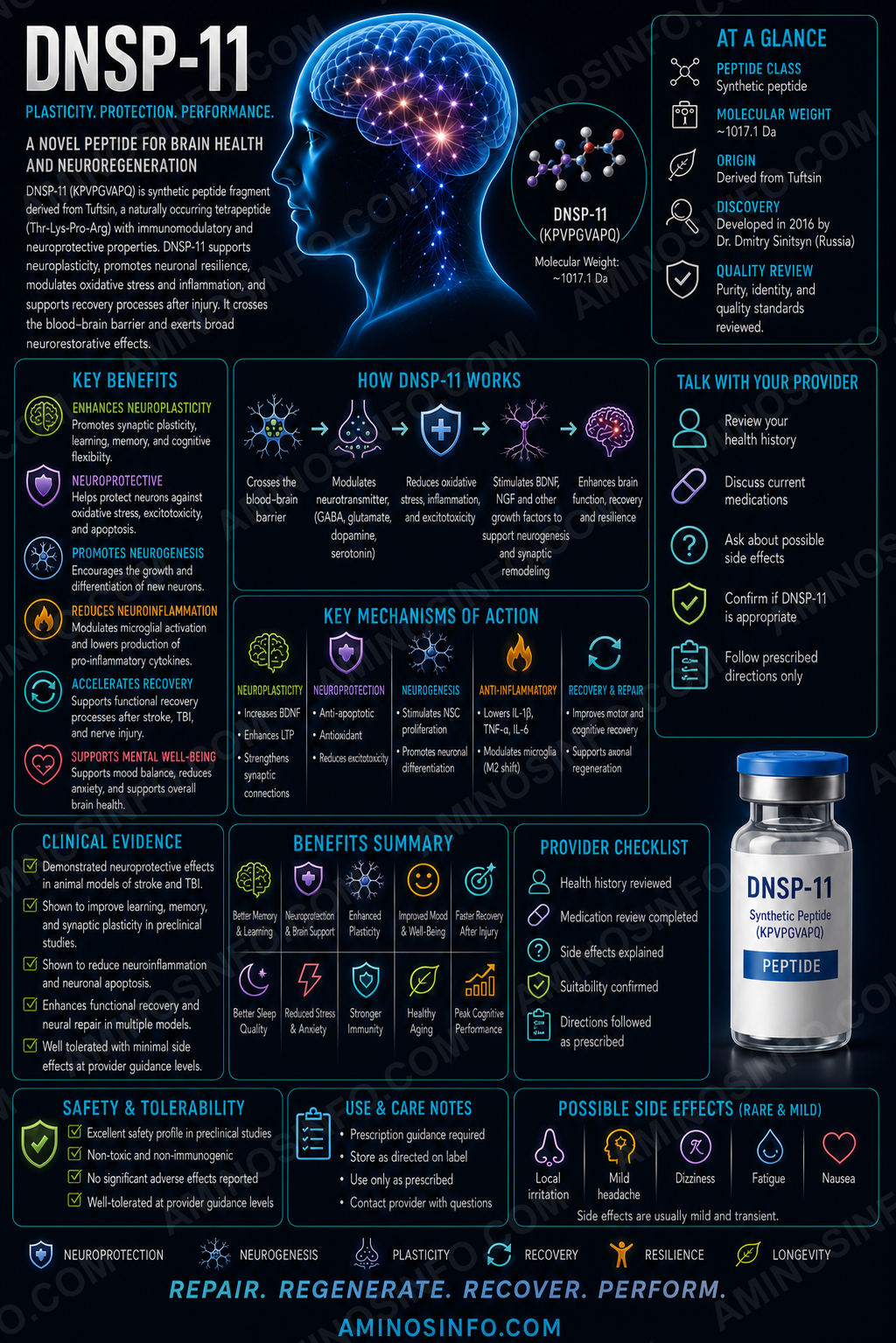
DNSP-11 stands for Dopamine Neuron Stimulating Peptide-11. It is an investigational 11-amino-acid amidated peptide predicted to be produced from the pro-domain of human glial cell line-derived neurotrophic factor (proGDNF).
Researchers developed DNSP-11 as a smaller, easier-to-manufacture alternative to full-length GDNF. It produces several GDNF-like outcomes in dopaminergic models, including neuronal survival, neurite growth, increased dopamine function, and behavioral improvement after a 6-hydroxydopamine lesion. However, its signaling appears mechanistically distinct from GDNF.
Dopamine Neuron Stimulating Peptide-11
PPEAPAEDRSL-NH₂
11 amino acids
Human proGDNF
Dopaminergic neuroprotection
No
🧬 Molecular Structure
🧪 Published amino-acid sequence
Pro-Pro-Glu-Ala-Pro-Ala-Glu-Asp-Arg-Ser-Leu-NH₂
PPEAPAEDRSL-NH₂
Terminal chemistry
- Free N-terminal proline
- C-terminal leucinamide
- No cysteine residues
- No disulfide bonds
- Linear peptide
⚛️ Molecular Weight and 🧫 Formula
| Molecular formula | C50H81N15O18 |
|---|---|
| Average molecular weight | Approximately 1,180.3 g/mol |
| Peptide length | 11 amino acids |
| C-terminal modification | Amidated |
| Disulfide bonds | None |
| Typical research formulation | Acidic citrate-buffered solution in foundational studies |
Biotinylated DNSP-11
Some mechanistic studies used N-terminally biotinylated DNSP-11 for uptake and protein-interaction experiments. Biotin-DNSP-11 is a different analytical reagent with a higher molecular weight and should not be confused with the unmodified reference peptide.
📅 Discovery Timeline and Research History
1990s: GDNF characterized
GDNF was established as a potent survival and restorative factor for midbrain dopamine neurons, prompting extensive Parkinson’s disease research.
2000s: ProGDNF processing hypothesis
Sequence analysis identified dibasic cleavage sites in the GDNF pro-domain that could theoretically release smaller biologically active peptides.
2010: DNSP-11 foundational paper
The 11-residue amidated peptide was shown to increase dopaminergic-neuron survival and neurite outgrowth, protect cultured cells, increase dopamine and metabolites, and improve rotational behavior in 6-OHDA-lesioned rats.
2011: Physical and cellular characterization
DNSP-11, DNSP-5, and DNSP-17 were evaluated for solubility, secondary structure, cytoprotection, and resistance to selected experimental conditions.
2014: Dynamic dopamine-function study
A single nigral treatment produced time- and region-dependent changes in evoked striatal dopamine release and activated ERK1/2 in a dopaminergic cell line.
2015: Repeated intranasal rat delivery
Researchers developed a repeated intranasal method and reported increased dopamine turnover in the striatum and substantia nigra at one tested dose.
2018: Awake rhesus-macaque intranasal methodology
Repeated dose-escalation was evaluated over ten weeks, including tolerability, brain distribution, and dopamine-system target engagement.
2019: Rat-brain immunoreactivity mapping
DNSP-11 antibody signal was reported in substantia nigra, ventral tegmental area, hippocampus, cerebellum, and other regions, although antibodies could not definitively distinguish free peptide from larger precursor-containing material.
2022–2023: Continued cellular and aggregation research
Additional studies examined protection of human dopaminergic cells and inhibition of amyloid or IAPP aggregation.
Current status
No established phase 1–3 human clinical program or FDA-approved DNSP-11 product exists.
GDNF Precursor Origin and Proposed Processing
PreproGDNF
GDNF is synthesized as a larger precursor containing an N-terminal signal peptide, a pro-domain, and the mature GDNF growth-factor domain.
Dibasic cleavage sites
The human pro-domain contains predicted endoprotease-recognition sites that could release the DNSP-11 sequence.
C-terminal amidation
Published models propose additional processing to convert the C-terminal leucine into leucinamide, producing PPEAPAEDRSL-NH₂.
Rat homolog: brain excitatory peptide
A homologous rat proGDNF-derived peptide called brain excitatory peptide was reported to increase hippocampal neuronal excitability.
Endogenous existence remains partly unresolved
Immunoreactivity supports the presence of DNSP-11-containing sequences in brain tissue, but definitive mass-spectrometric demonstration of a naturally processed free 11-mer in humans remains limited.
🧠 Proposed Mechanism of Action
1. Noncanonical GDNF-like signaling
DNSP-11 produces neurotrophic outcomes similar to GDNF but does not appear to bind GFRα1 directly or activate the classic GFRα1–RET pathway.
2. ERK1/2 activation
A single treatment increased ERK1/2 phosphorylation in MN9D dopaminergic cells, suggesting a growth- and plasticity-related signaling mechanism.
3. Mitochondrial protection
DNSP-11 prevented stress-induced cytochrome-c release from mitochondria and reduced apoptosis in dopaminergic cell models.
4. Metabolic-protein interactions
Pull-down studies identified multiple cytosolic proteins, many involved in metabolism, including GAPDH. The direct functional relevance of each interaction remains uncertain.
5. Neuronal uptake
Injected DNSP-11 was rapidly detected in neuronal cytoplasm, nuclei, and neurites in the substantia nigra.
🎯 Target and Pathway Profile
| Target or pathway | Evidence status |
|---|---|
| GFRα1 | Direct binding was not supported in foundational studies. |
| RET | Canonical RET activation is not established. |
| ERK1/2 | Phosphorylation increased in dopaminergic cells. |
| Mitochondrial cytochrome c | Stress-induced release was reduced. |
| Caspase-3 and caspase-3/7 | Activation decreased in toxin-exposed cells. |
| GAPDH and metabolic proteins | Identified in pull-down experiments; significance remains exploratory. |
| Dopamine synthesis and turnover | Increased in several rodent studies. |
| Single validated receptor | Not identified. |
Dopamine-Neuron and Neurochemical Research
Primary mesencephalic neurons
DNSP-11 increased survival of tyrosine-hydroxylase-positive fetal rat midbrain neurons and enhanced neurite length and branching.
Normal adult rat substantia nigra
A single nigral administration increased resting striatal dopamine, DOPAC, and HVA approximately four weeks later in the foundational study.
Evoked dopamine release
Later work reported time- and striatal-subregion-dependent changes in potassium-evoked dopamine release, while some microdialysis measures were unchanged.
Dopamine turnover after intranasal dosing
Repeated intranasal administration increased dopamine turnover in the striatum and substantia nigra at an intermediate tested dose in normal rats.
Not a dopamine-receptor agonist
DNSP-11 does not act like levodopa, apomorphine, pramipexole, ropinirole, or other direct dopaminergic medicines.
Mitochondrial and Anti-Apoptotic Research
6-Hydroxydopamine
DNSP-11 reduced TUNEL-positive cells and caspase-3 activity after 6-OHDA exposure in dopaminergic cells.
Staurosporine
DNSP-11 protected nutrient-deprived B65 cells against staurosporine-induced cytotoxicity, whereas GDNF did not protect in that specific assay.
Gramicidin
Protection against gramicidin-related mitochondrial and ionic stress was also reported.
3-Nitropropionic acid
Later work reported dose-dependent protection from mitochondrial-complex-II-related toxicity.
Mitochondrial mechanism
Preservation of cytochrome c within mitochondria supports an anti-apoptotic mechanism upstream of caspase activation.
Survival signaling tradeoff
Broad anti-apoptotic activity may protect vulnerable neurons, but long-term effects on damaged, senescent, or malignant cells have not been adequately evaluated.
Parkinson’s Disease-Model Research
6-OHDA rat model
Unilateral 6-hydroxydopamine lesions damage nigrostriatal dopamine neurons and produce asymmetric motor behavior.
Rotational behavior
A single DNSP-11 injection into the lesioned substantia nigra reduced apomorphine-induced rotations by approximately half, beginning one week after treatment and persisting for several weeks.
Nigral dopamine and DOPAC
DNSP-11 increased dopamine and DOPAC tissue content in the lesioned substantia nigra.
Restorative interpretation
These results suggest improved function of surviving neurons rather than proven replacement of neurons lost to advanced disease.
Model limitations
The 6-OHDA model is an acute toxin lesion and does not fully reproduce alpha-synuclein pathology, progressive neuronal loss, nonmotor symptoms, or human Parkinson’s disease heterogeneity.
No human disease-modifying evidence
DNSP-11 has not been shown to slow progression, improve Unified Parkinson’s Disease Rating Scale scores, reduce falls, or alter biomarkers in humans.
Intranasal Delivery Research
Rationale
Intranasal administration is intended to bypass some blood–brain barrier limitations through olfactory and trigeminal pathways.
Normal-rat study
Repeated intranasal DNSP-11 was administered five days per week for three weeks. One dose increased dopamine turnover in the striatum and substantia nigra.
Lesion studies
Dissertation and preclinical work examined intranasal DNSP-11 before or after 6-OHDA lesions, with selected protective and neurochemical findings.
Dose-response complexity
The intermediate dose produced effects that were not necessarily larger at the highest dose, highlighting a potentially non-linear response.
Human translation limitations
Nasal anatomy, mucociliary clearance, peptidases, device performance, formulation, and brain distribution differ substantially between rodents and humans.
Nonhuman-Primate Research
Awake dosing methodology
A repeated intranasal administration method was developed for awake rhesus macaques without routine sedation.
Ten-week dose escalation
Animals received escalating DNSP-11 exposure while investigators evaluated tolerability, dopamine-related target engagement, and nasal administration feasibility.
Radiolabeled distribution
A modified iodine-125-labeled DNSP-11 tracer was used to examine distribution after intranasal delivery.
Tolerability
The methodology study reported general feasibility and no major overt toxicity signal under the tested conditions.
Limitations
The study was not a human efficacy trial, used a modified tracer for distribution analysis, involved few animals, and did not establish disease modification.
Endogenous Peptide and Immunoreactivity Evidence
Postnatal rat substantia nigra
DNSP-11 antibody staining co-localized with tyrosine-hydroxylase-positive neurons in young rats.
Adult rat-brain mapping
Immunoreactivity was reported in substantia nigra, ventral tegmental area, dentate gyrus, cerebellum, locus coeruleus, and other regions.
Antibody limitation
Polyclonal antibodies may detect the DNSP-11 sequence within proGDNF or other precursor fragments and cannot alone prove the presence of the free amidated 11-mer.
Mass-spectrometric confirmation
Definitive endogenous identification would require targeted high-resolution mass spectrometry that distinguishes the processed amidated peptide from larger proteins and homologous fragments.
Protein-Aggregation Research
IAPP and amyloid-beta
A 2023 study reported that DNSP-11 was nonaggregating and inhibited aggregation of human islet amyloid polypeptide and amyloid-beta 42 in experimental assays.
Potential relevance
This raises interest in diabetes- and Alzheimer’s-related protein aggregation, but it is separate from the dopaminergic neurotrophic literature.
Mechanism uncertain
Direct binding, steric inhibition, altered nucleation, or assay-specific effects require further study.
No clinical anti-amyloid evidence
DNSP-11 has not been shown to reduce pancreatic amyloid, brain amyloid PET signal, cognitive decline, or diabetes complications in humans.
Evidence Limitations and Clinical Interpretation
Preclinical evidence only
No established controlled human trial has demonstrated safety, pharmacokinetics, or efficacy.
Concentrated research program
Much of the DNSP-11 literature comes from connected University of Kentucky research groups.
Acute toxin models
6-OHDA and staurosporine models capture selected injury mechanisms but not the full biology of idiopathic Parkinson’s disease.
Unknown direct receptor
The lack of a validated receptor complicates potency testing, dose selection, off-target screening, and biomarker development.
Endogenous processing remains incompletely proven
Sequence prediction and immunoreactivity do not fully establish physiological production of the free human peptide.
Behavioral and biochemical effects can diverge
Not all studies found consistent changes across dopamine-release methods, locomotion, or brain regions.
Safety and Regulatory Considerations
No established human safety profile
No approved label defines dosage, route, contraindications, interactions, pregnancy safety, or long-term adverse effects.
Potential dopaminergic risks
- Dyskinesia or abnormal movements
- Impulsivity or compulsive behavior
- Sleep disturbance
- Psychosis or hallucination risk in susceptible individuals
- Blood-pressure or autonomic changes
- Interaction with levodopa or dopamine agonists
Potential survival-signaling risks
Long-term anti-apoptotic and ERK activation could have unintended effects on abnormal cells, tumors, or maladaptive plasticity.
Intranasal risks
Repeated nasal delivery may cause irritation, mucosal injury, altered smell, infection risk, or variable dosing.
Product-quality risk
Unapproved material may contain the wrong sequence, nonamidated peptide, deletion products, aspartimide or isoaspartate, oxidized residues, endotoxin, residual solvents, or inaccurate net content.
Regulatory status
DNSP-11 is not FDA approved.
🧪 Laboratory Testing Methods
| Method | Purpose | Important limitation |
|---|---|---|
| RP-HPLC / UPLC | Separates DNSP-11 from deletion peptides, nonamidated material, and degradants. | Area purity does not prove identity or net content. |
| LC-HRMS | Confirms intact mass and elemental composition. | Sequence isomers may share mass. |
| MS/MS sequencing | Confirms PPEAPAEDRSL residue order and C-terminal amidation. | Leucine/isoleucine differentiation requires care. |
| Edman degradation | Orthogonally confirms N-terminal sequence. | Does not verify the C-terminal amide well. |
| C-terminal amidation assay | Distinguishes DNSP-11 from PPEAPAEDRSL-OH. | Requires targeted high-resolution analysis. |
| Amino-acid analysis | Confirms composition and supports net-content measurement. | Does not prove sequence order. |
| Chiral amino-acid analysis | Detects D-amino-acid epimers. | Hydrolysis may introduce artifacts. |
| Aspartimide and isoaspartate assay | Detects synthesis and storage-related Asp degradation. | Requires specialized chromatography or MS. |
| Net peptide-content assay | Measures actual DNSP-11 quantity. | Must correct for water, counterions, and residual solvents. |
| ERK phosphorylation assay | Measures one reported functional response. | No universally validated release-potency assay exists. |
| 6-OHDA cell-protection assay | Measures dopaminergic-cell protection. | Results depend strongly on cell line and toxin conditions. |
| Cytochrome-c and caspase assay | Evaluates mitochondrial anti-apoptotic activity. | Not specific to DNSP-11 identity. |
| GFRα1/RET counter-test | Confirms that the product does not merely behave as contaminating GDNF. | Negative testing does not identify the true receptor. |
| Plasma, nasal-fluid, and protease stability | Measures degradation and metabolite formation. | Animal matrices do not fully predict humans. |
| Brain/plasma pharmacokinetics | Measures exposure and CNS distribution. | Human pharmacokinetics remain unknown. |
| Microbial limits, sterility, and endotoxin | Evaluate route-specific microbiological quality. | Requirements depend on the final dosage form. |
| Stability-indicating assay | Tracks deamidation, aspartimide, hydrolysis, aggregation, and potency. | Requires defined formulation and qualified standards. |
📄 How to Interpret a DNSP-11 COA
- Verify the exact published sequence: PPEAPAEDRSL-NH₂.
- Confirm that it contains 11 residues: conflicting vendor sequences should be rejected.
- Confirm C-terminal amidation: the free-acid peptide is not the published DNSP-11 reference compound.
- Confirm formula and molecular weight: C₅₀H₈₁N₁₅O₁₈ and approximately 1,180.3 g/mol.
- Use MS/MS and an orthogonal sequence method: HPLC and intact mass alone are insufficient.
- Review deletion sequences, nonamidated peptide, aspartimide, isoaspartate, epimers, aggregates, water, salts, and residual solvents.
- Measure net peptide content: “99% purity” is not the same as the labeled milligram amount.
- Require a relevant functional assay: ERK activation or dopaminergic-cell protection may be used, but no validated clinical potency assay exists.
- Match microbiological testing to route: intranasal and injectable finished products require route-specific controls.
- Do not infer efficacy: a COA cannot prove dopamine restoration, brain delivery, Parkinson’s treatment, neuroprotection, or human safety.
📊 DNSP-11 vs GDNF vs Neurturin
| Feature | DNSP-11 | GDNF | Neurturin |
|---|---|---|---|
| Type | 11-residue propeptide | Full neurotrophic protein | GDNF-family protein |
| Main receptor | Unknown | GFRα1–RET | GFRα2–RET |
| Molecular size | ~1.18 kDa | Much larger dimeric protein | Much larger protein |
| Main research theme | Dopaminergic neuroprotection and metabolism | Dopamine-neuron survival and restoration | Dopamine-neuron trophic support |
| FDA approved? | No | No for Parkinson’s disease | No |
DNSP-11 vs DNSP-5 vs DNSP-17
| Peptide | Length | Source region | Research emphasis |
|---|---|---|---|
| DNSP-11 | 11 amino acids | GDNF pro-domain | Dopamine function, mitochondria, Parkinson’s models |
| DNSP-5 | 5 amino acids | Predicted GDNF-related processing product | Dopamine differentiation and cytoprotection |
| DNSP-17 | 17 amino acids | Predicted GDNF-related sequence | Physical and cellular characterization |
DNSP-11 vs P21 vs FGL vs Dihexa
| Compound | Main proposed pathway | Main research theme |
|---|---|---|
| DNSP-11 | Unknown receptor; ERK and mitochondrial protection | Dopaminergic-neuron support |
| P021 | LIF/BDNF/GSK3β | Neurogenesis, tau, cognition |
| FGL | NCAM–FGFR1 | Synaptic plasticity and memory |
| Dihexa | HGF/c-Met | Synaptogenesis |
DNSP-11 vs Evidence-Based Parkinson’s Care
| Approach | Established role | Difference from DNSP-11 |
|---|---|---|
| Levodopa | Most effective symptomatic motor treatment | Approved, defined dosing and risks |
| Dopamine agonists | Symptomatic treatment in selected patients | Direct receptor pharmacology and human evidence |
| MAO-B and COMT inhibitors | Extend dopaminergic therapy effects | Approved human medicines |
| Deep-brain stimulation | Selected advanced motor complications | Established surgical therapy |
| DNSP-11 | Experimental trophic peptide | No proven human safety or efficacy |
🔗 Related Peptides and Pathways
- GDNF: Parent neurotrophic protein.
- DNSP-5 and DNSP-17: Related predicted GDNF-derived peptides.
- Brain excitatory peptide: Rat homolog associated with synaptic excitability.
- ERK1/2: Signaling pathway activated in dopaminergic cells.
- Cytochrome c and caspases: Mitochondrial apoptosis pathways inhibited in cell research.
- GAPDH: Metabolic protein identified in pull-down studies.
- GFRα1 and RET: Canonical GDNF receptors not directly activated by DNSP-11 in foundational work.
- 6-OHDA: Dopaminergic neurotoxin used in Parkinson’s models.
🖼️ Original Diagram Specifications
Diagram 1: DNSP-11 sequence
Show PPEAPAEDRSL-NH₂ with the C-terminal amide highlighted and acidic, basic, polar, and hydrophobic residues labeled.
Diagram 2: PreproGDNF processing
Show signal peptide, pro-domain, predicted dibasic cleavage sites, DNSP-11, and mature GDNF.
Diagram 3: GDNF versus DNSP-11 signaling
Show GDNF activating GFRα1–RET and DNSP-11 following an unidentified receptor or intracellular pathway toward ERK and mitochondria.
Diagram 4: Mitochondrial protection
Show toxin exposure, mitochondrial depolarization, cytochrome-c release, caspase activation, and DNSP-11 inhibition.
Diagram 5: Nigrostriatal pathway
Show substantia nigra dopamine neurons, striatal projections, dopamine release, 6-OHDA lesion, and DNSP-11-associated functional changes.
Diagram 6: Intranasal delivery
Show nasal cavity, olfactory and trigeminal routes, brain distribution, degradation barriers, and target regions.
Diagram 7: COA workflow
Show exact sequence, amidation, HRMS, MS/MS, Edman analysis, Asp degradation, net content, functional assay, microbiology, and stability.
❓ Frequently Asked Questions
Is DNSP-11 a peptide?
Yes. It is a synthetic 11-amino-acid amidated peptide predicted from the human GDNF pro-domain.
What is its exact sequence?
PPEAPAEDRSL-NH₂.
What is its molecular formula?
C₅₀H₈₁N₁₅O₁₈.
What is its molecular weight?
Approximately 1,180.3 g/mol.
Is DNSP-11 the same as GDNF?
No. It is a short pro-domain-derived peptide with different receptor biology.
Does DNSP-11 bind GFRα1?
Foundational studies did not detect direct interaction with GFRα1.
What is DNSP-11 studied for?
Dopaminergic-neuron survival, mitochondrial protection, dopamine function, Parkinson’s models, and intranasal brain delivery.
Does DNSP-11 increase dopamine?
Several rodent studies reported increased dopamine, dopamine metabolites, turnover, or evoked release in selected conditions.
Does it regrow dopamine neurons?
It improved survival and neurite growth in cells and function of surviving neurons in animals, but proven regeneration of human neurons has not been established.
Is DNSP-11 FDA approved?
No.
Has it been tested in humans?
No established controlled human clinical trial has demonstrated safety or efficacy.
Can DNSP-11 be administered intranasally?
Intranasal delivery has been studied in rats and rhesus macaques, but no approved human protocol exists.
Does it treat Parkinson’s disease?
No human therapeutic benefit has been established.
Is DNSP-11 naturally produced in the brain?
Immunoreactivity and sequence predictions support the hypothesis, but definitive endogenous free-peptide identification remains incomplete.
Does 99% HPLC purity prove DNSP-11 identity?
No. Exact sequence, amidation, mass, degradation products, net content, and functional activity require separate confirmation.
Final Thoughts
DNSP-11 is a legitimate 11-residue GDNF-prodomain-derived research peptide with a focused preclinical literature in dopaminergic neurobiology. Its published sequence is PPEAPAEDRSL-NH₂, and its effects differ mechanistically from canonical GDNF signaling.
Cell and rodent studies report improved dopaminergic-neuron survival, neurite growth, mitochondrial protection, dopamine function, and motor behavior after neurotoxic injury. Repeated intranasal delivery has also been investigated in rats and rhesus macaques. However, no human efficacy or long-term safety evidence establishes DNSP-11 as a Parkinson’s treatment.
Legitimate material should be tested for exact sequence, C-terminal amidation, molecular mass, deletion peptides, aspartimide and isoaspartate, stereochemical integrity, net peptide content, functional activity, route-specific microbiological quality, and stability.
📚 References
- Bradley LH, et al. Dopamine Neuron Stimulating Actions of a GDNF Propeptide. PLoS ONE. 2010.
- Kelps KA, et al. Evaluation of the Physical and In Vitro Protective Activity of Three Synthetic Peptides Derived from the Pro- and Mature GDNF Sequence. 2011.
- Fuqua JL, et al. Dynamic Changes in Dopamine Neuron Function after DNSP-11 Treatment. Peptides. 2014.
- Stenslik MJ, et al. Methodology and Effects of Repeated Intranasal Delivery of DNSP-11 in a Rat Model of Parkinson’s Disease. Journal of Neuroscience Methods. 2015.
- Stenslik MJ, et al. Methodology and Effects of Repeated Intranasal Delivery of DNSP-11 in Rhesus Macaques. Journal of Neuroscience Methods. 2018.
- Sonne JWH, et al. Spatial and temporal immunoreactivity in the rat brain using a DNSP-11 antibody. Peptides. 2019.
- Parmar MS, et al. DNSP-11 reduces 6-OHDA-induced caspase activation in human dopaminergic cells. FASEB Journal. 2022.
- Liu QR, et al. Novel hominid-specific IAPP isoforms and inhibition of IAPP and Aβ aggregation by DNSP-11. 2023.
- Immonen T, et al. Brain excitatory peptide, a rat proGDNF-derived peptide, increases synaptic excitability. Neuroscience research literature.
- Lin LF, et al. GDNF: a glial cell line-derived neurotrophic factor for midbrain dopaminergic neurons. Science. 1993.
- Airaksinen MS, Saarma M. The GDNF family: signalling, biological functions and therapeutic value. Nature Reviews Neuroscience.
- Ibáñez CF, Andressoo JO. Biology of GDNF and its receptors. Neurobiology of Disease.
- Sidorova YA, Saarma M. Can growth factors cure Parkinson’s disease? Trends in Pharmacological Sciences.
- Sidorova YA, et al. Small molecules and peptides targeting GDNF-family receptor pathways. International Journal of Molecular Sciences. 2020.
- Whone AL, et al. Randomized trial of intermittent intraputamenal GDNF in Parkinson’s disease. Brain.
- Gill SS, et al. Direct brain infusion of GDNF in Parkinson disease. Nature Medicine.
- Lang AE, et al. Randomized controlled trial of intraputamenal GDNF infusion. Annals of Neurology.
- Nutt JG, et al. Randomized trial of glial cell line-derived neurotrophic factor. Neurology.
- Marks WJ, et al. Gene delivery of neurturin for Parkinson’s disease. Lancet Neurology.
- Marks WJ, et al. Safety and tolerability of CERE-120 in Parkinson’s disease. Lancet Neurology.
- Kordower JH, et al. Clinicopathological findings following neurturin gene therapy. Annals of Neurology.
- Dauer W, Przedborski S. Parkinson’s disease: mechanisms and models. Neuron.
- Kalia LV, Lang AE. Parkinson’s disease. Lancet.
- Bloem BR, Okun MS, Klein C. Parkinson’s disease. Lancet. 2021.
- Surmeier DJ, Obeso JA, Halliday GM. Selective neuronal vulnerability in Parkinson disease. Nature Reviews Neuroscience.
- Schober A. Classic toxin-induced animal models of Parkinson’s disease: 6-OHDA and MPTP. Cell and Tissue Research.
- Ungerstedt U. 6-Hydroxydopamine-induced degeneration of the nigrostriatal dopamine pathway. European Journal of Pharmacology.
- Deumens R, et al. Modeling Parkinson’s disease in rats: an evaluation of 6-OHDA lesions. Experimental Neurology.
- Bové J, Perier C. Neurotoxin-based models of Parkinson’s disease. Neuroscience.
- Burke RE, O’Malley K. Axon degeneration in Parkinson’s disease. Experimental Neurology.
- Exner N, et al. Mitochondrial dysfunction in Parkinson’s disease. EMBO Journal.
- Pickrell AM, Youle RJ. The roles of PINK1, parkin, and mitochondrial fidelity in Parkinson’s disease. Neuron.
- Green DR, Kroemer G. The pathophysiology of mitochondrial cell death. Science.
- Tait SWG, Green DR. Mitochondria and cell death. Cold Spring Harbor Perspectives in Biology.
- Garrido C, et al. Mechanisms of cytochrome c release from mitochondria. Cell Death and Differentiation.
- Elmore S. Apoptosis: a review of programmed cell death. Toxicologic Pathology.
- Ramos JW. The regulation of extracellular signal-regulated kinase in mammalian cells. International Journal of Biochemistry & Cell Biology.
- Roux PP, Blenis J. ERK and p38 MAPK-activated protein kinases. Microbiology and Molecular Biology Reviews.
- Chuang DM, et al. Neuroprotective effects of ERK signaling. Journal of Neurochemistry.
- Mazzola JL, Sirover MA. Subcellular alteration of GAPDH in neurodegenerative disease. Journal of Neuroscience Research.
- Butterfield DA, et al. GAPDH in neurodegenerative disorders. Journal of Alzheimer’s Disease.
- Illum L. Nasal drug delivery—possibilities, problems and solutions. Journal of Controlled Release.
- Lochhead JJ, Thorne RG. Intranasal delivery of biologics to the central nervous system. Advanced Drug Delivery Reviews.
- Dhuria SV, et al. Intranasal delivery to the central nervous system. Journal of Pharmaceutical Sciences.
- Hanson LR, Frey WH. Intranasal delivery bypasses the blood–brain barrier. Drug Delivery and Translational Research.
- Erdő F, et al. Evaluation of intranasal delivery route of drug administration for brain targeting. Brain Research Bulletin.
- Wen P, et al. Research progress on intranasal treatment for Parkinson’s disease. 2024.
- Fosgerau K, Hoffmann T. Peptide therapeutics: current status and future directions. Drug Discovery Today.
- Lau JL, Dunn MK. Therapeutic peptides: historical perspectives and current development trends. Bioorganic & Medicinal Chemistry.
- Craik DJ, Fairlie DP, Liras S, Price D. The future of peptide-based drugs. Chemical Biology & Drug Design.
- International Council for Harmonisation. ICH Q2(R2): Validation of Analytical Procedures.
- International Council for Harmonisation. ICH Q3A and Q3B: Impurities in New Drug Substances and Products.
- International Council for Harmonisation. ICH Q3C: Residual Solvents.
- International Council for Harmonisation. ICH Q1A(R2): Stability Testing of New Drug Substances and Products.
- International Council for Harmonisation. ICH M10: Bioanalytical Method Validation.
- United States Pharmacopeia General Chapter <621>: Chromatography.
- United States Pharmacopeia General Chapter <71>: Sterility Tests.
- United States Pharmacopeia General Chapter <85>: Bacterial Endotoxins Test.
- United States Pharmacopeia General Chapters <232> and <233>: Elemental Impurities.
Identity, sequence, proGDNF origin, dopaminergic, mitochondrial, Parkinson’s-model, intranasal, primate, aggregation, safety, and analytical evidence were reviewed in July 2026. DNSP-11 remains an unapproved investigational peptide.