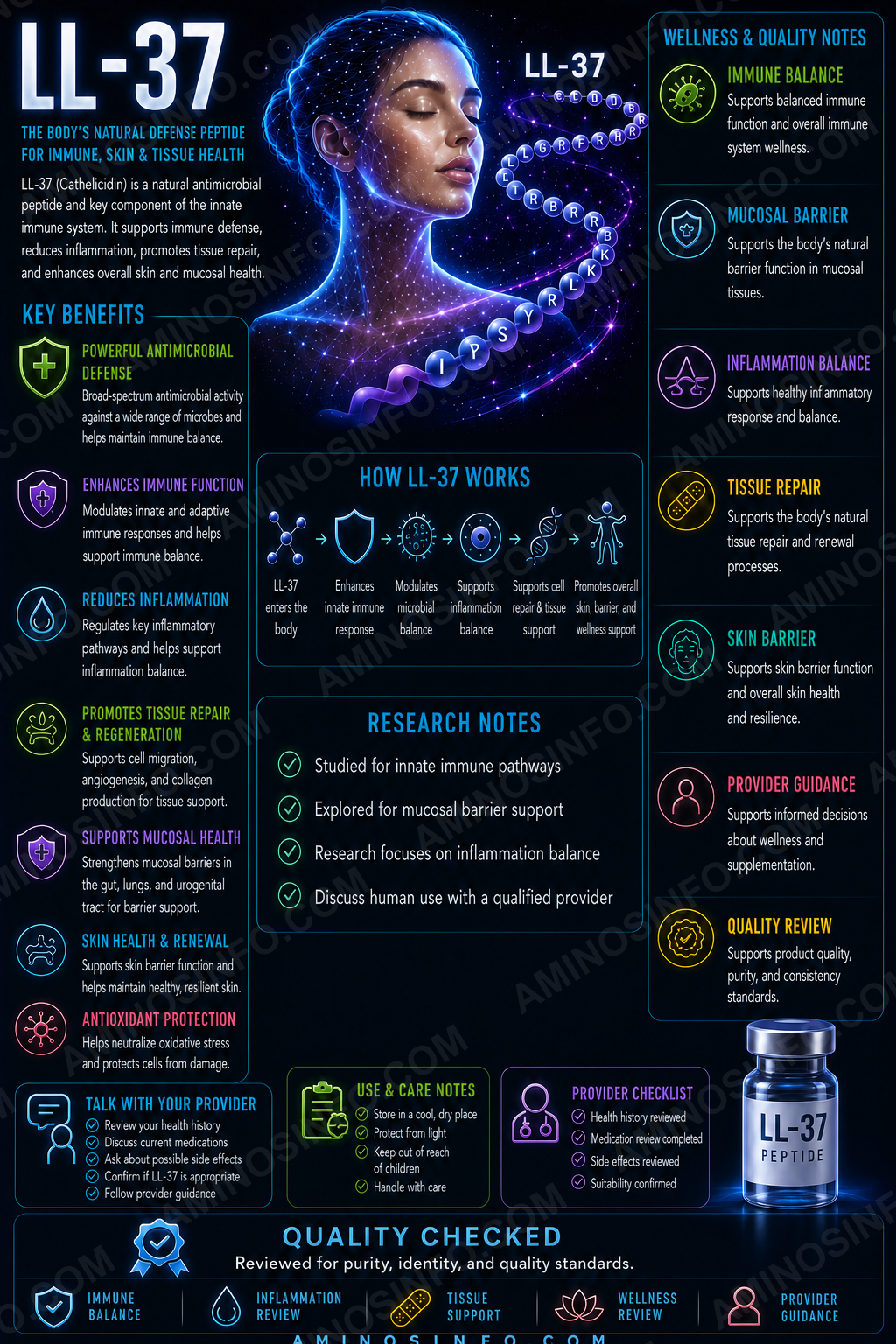
LL-37 (Human Cathelicidin): What It Is, How It Works, Benefits, and Research Overview
A comprehensive, evidence-graded review of LL-37, the 37-amino-acid human cathelicidin host-defense peptide involved in antimicrobial activity, immune regulation, chemotaxis, biofilm biology, angiogenesis, and tissue repair.
What Is LL-37?
LL-37 is the only known human cathelicidin antimicrobial peptide. It is generated by proteolytic cleavage of the precursor protein hCAP18, encoded by the CAMP gene.
The name comes from its first two amino acids—leucine and leucine—and its length of 37 amino-acid residues.
Human host-defense peptide
hCAP18
CAMP
37 amino acids
Approximately +6 at physiologic pH
No synthetic LL-37 drug approval
Major biological roles
- Direct microbial membrane disruption
- Neutralization and binding of bacterial products such as LPS
- Immune-cell recruitment and chemotaxis
- Regulation of inflammatory signaling
- Biofilm inhibition
- Keratinocyte and fibroblast migration
- Angiogenesis and wound repair
- Interaction with nucleic acids and pattern-recognition receptors
🧬 Structure, Sequence, and Molecular Properties
🧪 Amino-acid sequence
LLGDFFRKSKEKIGKEFKRIVQRIKDFLRNLVPRTES
Structural characteristics
- 37 amino-acid residues
- Linear peptide with no disulfide bridge
- Strongly cationic because of multiple lysine and arginine residues
- Amphipathic, meaning hydrophobic and positively charged surfaces segregate when the peptide forms an alpha helix
- Can form monomers, dimers, tetramers, and other oligomeric assemblies depending on concentration and environment
- Structure changes in water, salt solutions, membranes, and microbial surfaces
⚛️ Molecular properties
| Representative molecular formula | C205H340N60O53 |
|---|---|
| Average molecular weight | Approximately 4493.3 Da |
| Monoisotopic mass | Approximately 4490.8 Da |
| Length | 37 residues |
| Net charge | Approximately +6 at physiologic pH |
| Disulfide bonds | None |
| Dominant membrane-associated structure | Amphipathic alpha helix |
How Does the Body Produce LL-37?
hCAP18 precursor
LL-37 is stored as part of the inactive precursor hCAP18. Cleavage releases the active C-terminal LL-37 peptide.
Major sources
- Neutrophils
- Monocytes and macrophages
- Keratinocytes
- Airway epithelial cells
- Gastrointestinal epithelial cells
- Salivary and reproductive tissues
Regulation
Expression is influenced by infection, inflammation, tissue injury, cytokines, and vitamin-D-receptor signaling.
Proteolytic processing
Different tissues can produce LL-37 fragments with altered antimicrobial, inflammatory, and cytotoxic properties.
📅 Discovery and Research Timeline
- 1990s: Human cathelicidin hCAP18 and its active C-terminal peptide LL-37 were characterized.
- 2000s: Research expanded from antimicrobial activity to chemotaxis, receptor signaling, wound repair, and biofilm biology.
- 2010s: LL-37 became linked to psoriasis, rosacea, lupus-related nucleic-acid signaling, angiogenesis, and tissue-dependent cancer effects.
- 2014: A small randomized clinical study reported that topical LL-37 was generally well tolerated and improved healing predictors in chronic venous leg ulcers at lower tested doses.
- 2014 onward: Intratumoral LL-37 was evaluated in an early melanoma study to explore immune stimulation and dose selection.
- 2020s: Research increasingly focuses on delivery systems, stable analogues, nanoparticles, wound dressings, antimicrobial combinations, and reducing host-cell toxicity.
- Current status: No FDA-approved synthetic LL-37 therapeutic exists.
🧠 How Does LL-37 Work?
1. Electrostatic membrane binding
Bacterial membranes contain negatively charged phospholipids, lipopolysaccharide, and lipoteichoic acid. Positively charged LL-37 is attracted to these surfaces.
2. Membrane disruption
LL-37 inserts into microbial membranes and can create transient lesions or pores, disturb lipid organization, and cause leakage of cellular contents.
3. Intracellular microbial effects
After membrane access, LL-37 may interact with nucleic acids, proteins, and metabolic systems.
4. Toxin and endotoxin binding
LL-37 can bind LPS and other microbial components, changing how innate immune receptors respond.
5. Host receptor signaling
Reported signaling partners include FPR2, P2X7, EGFR transactivation pathways, and other membrane-associated systems. Effects depend heavily on cell type, concentration, and local environment.
6. Nucleic-acid transport
LL-37 can form complexes with self-DNA or RNA and deliver them to intracellular Toll-like receptors. This can strengthen antimicrobial sensing but also contribute to autoimmune inflammation.
Antibacterial, Antifungal, and Antiviral Research
Antibacterial activity
LL-37 has demonstrated activity against many Gram-positive and Gram-negative organisms in vitro, including resistant pathogens.
Antifungal activity
Experimental activity has been reported against Candida and other fungi.
Antiviral activity
LL-37 may bind viral envelopes, disrupt membranes, aggregate particles, alter viral entry, and regulate antiviral immune responses.
Physiologic-environment limitation
Activity often falls in the presence of salts, serum proteins, mucus, wound fluid, DNA, and proteases. Strong activity in a simple laboratory buffer may not translate to human tissue.
Host-cell toxicity
The same membrane-active properties that damage microbes can injure mammalian cells at sufficiently high concentrations.
Biofilm Research
Biofilm prevention
LL-37 can reduce microbial attachment, interfere with early biofilm formation, and alter bacterial signaling.
Established biofilms
Activity against mature biofilms is variable and often weaker than prevention of initial formation.
Combination strategies
Researchers study LL-37 with antibiotics, nanoparticles, hydrogels, wound dressings, and modified fragments to improve local activity.
Resistance considerations
Although membrane-active peptides attack multiple targets, bacteria can still resist LL-37 through surface-charge changes, proteases, efflux, capsules, and biofilm matrices.
Immune and Inflammatory Signaling
Chemotaxis
LL-37 can recruit neutrophils, monocytes, mast cells, T cells, and other immune populations.
Inflammation can increase or decrease
Depending on context, LL-37 can suppress excessive responses to microbial products or amplify cytokine and interferon pathways.
Innate-adaptive immune bridge
LL-37 influences dendritic-cell maturation, antigen presentation, leukocyte migration, and cytokine production.
Extracellular-DNA complexes
LL-37 protects nucleic acids from degradation and facilitates entry into immune cells, contributing to type-I-interferon signaling in psoriasis and lupus-related models.
Concentration and tissue determine effect
Low local concentrations may support repair and defense, while persistent or excessive expression may sustain inflammation.
Wound-Healing and Angiogenesis Research
Re-epithelialization
LL-37 promotes keratinocyte migration and may activate EGFR-related repair pathways.
Fibroblast activity
It can influence fibroblast migration, extracellular-matrix production, and wound contraction.
Angiogenesis
LL-37 promotes endothelial-cell responses and new blood-vessel formation through several signaling pathways.
Chronic venous leg ulcers
A small randomized topical study found acceptable tolerability and favorable healing-related effects at lower tested doses, while the highest tested exposure was less favorable.
Delivery challenge
Proteases in chronic wound fluid can degrade LL-37. Modern approaches use hydrogels, nanoparticles, dressings, peptide analogues, and sustained-release systems.
Skin Disease, Psoriasis, and Rosacea
Protective skin defense
LL-37 contributes to antimicrobial defense and barrier repair.
Psoriasis
LL-37 forms complexes with self-DNA and RNA, enabling activation of TLR9, TLR7, and TLR8 pathways and type-I-interferon signaling. High lesional LL-37 may therefore contribute to disease pathogenesis.
Rosacea
Abnormal processing and increased activity of cathelicidin peptides are implicated in inflammation, vascular changes, and lesion formation.
Atopic dermatitis
Reduced antimicrobial-peptide activity may contribute to infection susceptibility in some patients, although disease biology is complex.
Not a universal skin treatment
Because LL-37 can improve repair yet worsen inflammatory signaling, disease-specific context is essential.
Gut, Respiratory, and Systemic Research
Respiratory tract
LL-37 participates in airway antimicrobial defense, mucus biology, inflammatory signaling, and epithelial repair.
Gastrointestinal tract
Research includes barrier function, microbial ecology, inflammatory bowel disease, and epithelial repair.
Sepsis and endotoxin research
LL-37 can neutralize LPS but can also produce systemic toxicity depending on concentration and delivery.
Autoimmune disease
LL-37–nucleic-acid complexes are studied in systemic lupus erythematosus, psoriasis, and other interferon-driven disorders.
Vitamin D connection
Vitamin-D signaling can increase CAMP expression, linking endocrine status to innate immune defense.
Cancer Research: Protective and Tumor-Promoting Effects
Potential anticancer actions
LL-37 and selected fragments can disrupt tumor-cell membranes, induce apoptosis, modify immune responses, and enhance drug delivery in some experimental models.
Potential tumor-promoting actions
In other tissues, LL-37 may stimulate proliferation, angiogenesis, migration, inflammatory signaling, and recruitment of tumor-supportive cells.
Tissue dependence
Research has associated LL-37 with tumor promotion in several ovarian, breast, lung, pancreatic, prostate, melanoma, and squamous-cell models, while inhibitory effects have been reported in some gastric, colon, hematologic, and other systems.
Intratumoral melanoma study
An early clinical study investigated intratumoral LL-37 to determine dose and immune effects in melanoma. This does not establish a general anticancer indication.
No established oncology role
LL-37 is not an approved cancer treatment, immune adjuvant, or preventive therapy.
Human Clinical Research
Chronic leg ulcers
Topical LL-37 was evaluated in a small randomized controlled study. Lower tested doses appeared safe and were associated with improved healing predictors; the dose-response was not simply “more is better.”
Melanoma
An early intratumoral study explored safety, dose, and immune activation.
Biomarker research
Endogenous LL-37 has been measured in infections, inflammatory disorders, autoimmune disease, wounds, cardiovascular disease, and cancer.
What has not been established
- Safe systemic or injectable dose
- Long-term repeated-use safety
- Approved treatment for chronic infection
- Approved “immune boosting” use
- Validated oral or nasal systemic therapy
- Generalized wound-healing indication
Major Evidence Limitations
- Many antimicrobial results come from simplified in-vitro conditions
- Serum, salts, mucus, DNA, and proteases can reduce activity
- Host-cell toxicity overlaps with antimicrobial potency
- Human trials are small and route specific
- Endogenous-expression studies do not prove benefit from administering synthetic peptide
- Pro-inflammatory and anti-inflammatory effects vary by context
- Cancer effects are tissue dependent and sometimes opposing
- Peptide stability and delivery remain major obstacles
- Commercial research preparations may differ in salt, purity, aggregation, and biological potency
Potential Side Effects and Safety Considerations
No approved systemic safety profile
There is no validated systemic dose, injection schedule, therapeutic window, or long-term safety program for synthetic LL-37.
Potential direct toxicity
- Hemolysis
- Membrane damage to host cells
- Local pain, irritation, or tissue injury
- Inflammatory-cell activation
- Endothelial and epithelial toxicity at high exposure
Potential immune risks
- Immunogenicity and anti-drug antibodies
- Excess cytokine or interferon signaling
- Aggravation of psoriasis, rosacea, lupus-related pathways, or other inflammatory disease
- Unpredictable effects in immunocompromised patients
Angiogenesis and cancer concern
LL-37 can promote blood-vessel growth and proliferation in some contexts. This creates theoretical and experimentally supported concerns in certain cancers and proliferative disorders.
FDA compounding concern
FDA has specifically identified LL-37 as a bulk substance that may present significant safety risks because of immunogenicity, peptide-related impurities, and insufficient human safety information.
🧪 Laboratory Testing Methods
| Method | Purpose | Important limitation |
|---|---|---|
| RP-HPLC / UPLC | Separates intact LL-37 from deletion peptides, oxidation products, and synthesis impurities | Area purity does not prove sequence or active content |
| LC-HRMS | Confirms intact molecular mass near 4493.3 Da | Does not alone prove sequence or stereochemistry |
| MS/MS peptide mapping | Confirms all 37 residues and termini | Requires a qualified reference standard |
| Amino-acid analysis | Confirms composition and supports net peptide-content assignment | Does not prove residue order |
| Chiral amino-acid analysis | Detects epimerization or D-amino-acid contamination | Hydrolysis can introduce artifacts |
| Net peptide-content assay | Measures actual LL-37 mass | Must correct for TFA, acetate, water, and salts |
| Counterion assay | Measures TFA or acetate content | Gross powder weight can overstate peptide content |
| SEC-HPLC / DLS | Measures oligomers, aggregates, and particles | Oligomerization may be concentration and buffer dependent |
| Circular dichroism | Measures alpha-helical structure in membrane-like conditions | Structure varies strongly by environment |
| MIC / MBC assays | Measures antimicrobial inhibition and killing | Results depend on salt, serum, inoculum, and medium |
| Time-kill assay | Measures rate and extent of microbial killing | Does not establish human therapeutic exposure |
| Biofilm assays | Measure prevention or disruption of biofilms | Prevention and eradication are different endpoints |
| Hemolysis assay | Measures red-blood-cell membrane toxicity | Does not capture all host-cell toxicity |
| Mammalian cytotoxicity panel | Evaluates epithelial, endothelial, hepatic, renal, and immune-cell injury | Cell lines do not reproduce systemic exposure |
| LPS-binding / neutralization assay | Measures endotoxin interaction | Binding may not predict inflammatory effect in vivo |
| Chemotaxis and cytokine assays | Measure immune signaling | Highly cell-type and concentration dependent |
| Protease-stability assay | Measures degradation in serum, wound fluid, or tissue proteases | In-vitro stability may not predict tissue half-life |
| Endotoxin, sterility, and bioburden | Required for relevant research preparations | Research testing does not establish approved injectable safety |
| Stability-indicating assay | Tracks oxidation, hydrolysis, aggregation, adsorption, and potency loss | Requires validated forced-degradation studies |
📄 How to Interpret an LL-37 COA
- Verify the exact 37-residue sequence: LLGDFFRKSKEKIGKEFKRIVQRIKDFLRNLVPRTES.
- Confirm intact molecular mass near 4493.3 Da.
- Use MS/MS peptide mapping: HPLC plus intact mass is insufficient.
- Confirm L-amino-acid stereochemistry and screen for epimerization.
- State the salt form: TFA, acetate, or another counterion.
- Report net peptide content after correcting for water and counterions.
- Measure deletion peptides and oxidation products.
- Assess aggregation and concentration-dependent oligomerization.
- Test antimicrobial potency under physiologically relevant salt and serum conditions.
- Include hemolysis and mammalian-cell cytotoxicity.
- For wound research, include protease stability in wound fluid.
- For any parenteral research, require endotoxin, sterility, particles, and container-closure controls.
- Do not interpret a COA as proof of human safety, efficacy, or FDA equivalence.
📊 Comparison Tables
LL-37 vs Thymosin Alpha-1 vs KPV vs BPC-157
| Feature | LL-37 | Thymosin Alpha-1 | KPV | BPC-157 |
|---|---|---|---|---|
| Main focus | Host defense, antimicrobial and immune signaling | Immune modulation | Anti-inflammatory signaling | Tissue-protection research |
| Direct antimicrobial activity | High in vitro | Limited | Limited to moderate in models | Not primary |
| Inflammatory effect | Can increase or decrease | Immunomodulatory | Primarily anti-inflammatory research | Context dependent |
| FDA approved in U.S. | No | No | No | No |
LL-37 vs Human Defensins
| Feature | LL-37 | Alpha/Beta Defensins |
|---|---|---|
| Family | Cathelicidin | Defensin |
| Structure | Linear amphipathic helix | Disulfide-stabilized peptides |
| Human cathelicidin count | Only known human cathelicidin | Multiple human defensins |
| Main roles | Antimicrobial, chemotactic, repair, angiogenesis | Antimicrobial and immune signaling |
LL-37 vs Conventional Antibiotics
| Feature | LL-37 | Conventional antibiotic |
|---|---|---|
| Primary action | Membrane and multi-pathway host-defense effects | Defined microbial target or pathway |
| Immune effects | Extensive | Usually secondary |
| Physiologic stability | Limited by salts, proteins, and proteases | Drug dependent |
| Clinical approval | No synthetic LL-37 drug | Many approved agents |
Raw LL-37 vs Research-Qualified Material
| Attribute | Basic raw peptide | Research-qualified LL-37 |
|---|---|---|
| Identity | HPLC and mass claim | Sequence mapping, stereochemistry, termini, salt form |
| Potency | May be absent | MIC/MBC, biofilm, LPS, and immune assays |
| Safety testing | Often minimal | Hemolysis, cytotoxicity, endotoxin, aggregation |
| Net content | May use gross powder mass | Corrected for water and counterions |
| Human equivalence | Neither establishes an FDA-approved therapeutic product | |
🖼️ Original Diagram Specifications
- Peptide architecture: 37-residue sequence displayed as hydrophobic and positively charged faces of an alpha helix.
- Precursor processing: CAMP gene → hCAP18 → protease cleavage → LL-37.
- Microbial killing: Electrostatic binding, membrane insertion, pore formation, and leakage.
- Immune signaling: FPR2, P2X7, EGFR, TLR nucleic-acid complexes, chemotaxis, and cytokines.
- Wound repair: Keratinocyte migration, fibroblasts, angiogenesis, antimicrobial defense, and re-epithelialization.
- Benefit–risk balance: Host defense and healing opposite cytotoxicity, autoimmune inflammation, angiogenesis, and cancer complexity.
- COA workflow: Sequence, HRMS, peptide mapping, salt correction, aggregation, antimicrobial potency, hemolysis, sterility, and stability.
❓ Frequently Asked Questions
Is LL-37 a peptide?
Yes. It is a naturally occurring 37-amino-acid human host-defense peptide.
What does LL-37 stand for?
Its first two residues are leucine-leucine, and it contains 37 amino acids.
What is the sequence?
LLGDFFRKSKEKIGKEFKRIVQRIKDFLRNLVPRTES.
What is the molecular formula?
Approximately C₂₀₅H₃₄₀N₆₀O₅₃ for the parent peptide.
What is the molecular weight?
Approximately 4493.3 Da.
Is LL-37 FDA approved?
No synthetic LL-37 drug is FDA approved.
Is LL-37 an antibiotic?
It has direct antimicrobial activity but is more accurately described as a multifunctional host-defense peptide.
Does LL-37 break biofilms?
It can inhibit biofilm formation and sometimes affect established biofilms, but activity varies greatly by organism and conditions.
Does LL-37 promote wound healing?
Preclinical and limited human topical research suggests it can support re-epithelialization and repair, but no broad approved wound indication exists.
Can LL-37 worsen inflammation?
Yes. It can amplify nucleic-acid sensing and interferon pathways in psoriasis and other autoimmune contexts.
Is LL-37 anticancer?
Its effects are tissue dependent. It can suppress some tumor models and promote others.
Can LL-37 damage human cells?
Yes, especially at higher concentrations or under certain exposure conditions.
Does a high HPLC purity prove potency?
No. Physiologic antimicrobial activity, immune signaling, aggregation, cytotoxicity, and net peptide content must also be evaluated.
Can endogenous LL-37 research prove injected LL-37 is beneficial?
No. Natural local production and external systemic administration are biologically different.
Is there an established injectable dose?
No.
Final Thoughts
LL-37 is one of the most biologically complex antimicrobial peptides. It does much more than kill microbes: it recruits immune cells, binds microbial toxins and nucleic acids, regulates inflammation, affects angiogenesis, and participates in epithelial and wound repair.
This multifunctionality is also its central challenge. The same properties that support host defense can damage host membranes, intensify autoimmune signaling, encourage pathologic angiogenesis, or alter tumor behavior. Its effects depend on concentration, tissue, route, microbial environment, proteases, salts, serum proteins, and disease context.
Human topical wound research provides an important proof of concept, but synthetic LL-37 remains unapproved and lacks a validated systemic dose or long-term safety profile. FDA has specifically highlighted insufficient safety information, immunogenicity concerns, and peptide-characterization complexity for compounded LL-37.
Analytical verification requires more than a purity percentage. A credible evaluation must confirm the full 37-residue sequence, intact mass, stereochemistry, salt form, net peptide content, oxidation, truncations, aggregation, physiologically relevant antimicrobial potency, host-cell cytotoxicity, hemolysis, protease stability, endotoxin, and microbiological quality.
📚 References
- Voronko OE, et al. Antimicrobial Peptides of the Cathelicidin Family: Focus on LL-37. International Journal of Molecular Sciences. 2025.
- Kuroda K, Okumura K, Isogai H, Isogai E. The Human Cathelicidin Antimicrobial Peptide LL-37 and Mimics Are Potential Anticancer Drugs. Frontiers in Oncology. 2015.
- Ridyard KE, Overhage J. The Potential of Human Peptide LL-37 as an Antimicrobial Agent. Antibiotics. 2021.
- Xhindoli D, et al. The Human Cathelicidin LL-37—A Pore-Forming Antibacterial Peptide and Host-Cell Modulator. Biochimica et Biophysica Acta. 2016.
- Duplantier AJ, van Hoek ML. The Human Cathelicidin LL-37 as a Potential Treatment for Polymicrobial Infected Wounds. Frontiers in Immunology. 2013.
- Grönberg A, et al. Treatment with LL-37 Is Safe and Effective in Enhancing Healing of Hard-to-Heal Venous Leg Ulcers. Wound Repair and Regeneration. 2014.
- Morizane S, et al. Cathelicidin Antimicrobial Peptide LL-37 in Psoriasis Enables Keratinocyte Reactivity against TLR9 Ligands. Journal of Investigative Dermatology. 2012.
- Lande R, et al. Plasmacytoid Dendritic Cells Sense Self-DNA Coupled with Antimicrobial Peptide. Nature. 2007.
- Ganguly D, et al. Self-RNA–Antimicrobial Peptide Complexes Activate Human Dendritic Cells through TLR7 and TLR8. Journal of Experimental Medicine. 2009.
- Piktel E, et al. The Role of Cathelicidin LL-37 in Cancer Development. Archivum Immunologiae et Therapiae Experimentalis. 2016.
- Lu F, et al. Repurposing Human Antibacterial Peptide LL-37 for Cancer Treatment. Frontiers in Pharmacology. 2022.
- ClinicalTrials.gov. Intratumoral Injections of LL37 for Melanoma. NCT02225366.
- U.S. Food and Drug Administration. Certain Bulk Drug Substances for Use in Compounding That May Present Significant Safety Risks: Cathelicidin LL-37.
- Yang D, et al. LL-37, the Neutrophil Granule- and Epithelial Cell-Derived Cathelicidin, Utilizes FPRL1 as a Receptor to Chemoattract Human Peripheral Blood Neutrophils, Monocytes, and T Cells. Journal of Experimental Medicine. 2000.
- Tokumaru S, et al. Induction of Keratinocyte Migration via Transactivation of the EGFR by LL-37. Journal of Immunology. 2005.
- Koczulla R, et al. An Angiogenic Role for the Human Peptide Antibiotic LL-37/hCAP-18. Journal of Clinical Investigation. 2003.
- Salzer S, et al. Antimicrobial Peptide LL-37 and Angiogenesis Signaling. Arteriosclerosis, Thrombosis, and Vascular Biology.
- Overhage J, et al. Human Host Defense Peptide LL-37 Prevents Bacterial Biofilm Formation. Infection and Immunity. 2008.
- Turner J, et al. Activities of LL-37, a Cathelin-Associated Antimicrobial Peptide of Human Neutrophils. Antimicrobial Agents and Chemotherapy. 1998.
- Johansson J, et al. Conformation-Dependent Antibacterial Activity of LL-37. Journal of Biological Chemistry. 1998.
- Oren Z, et al. Structure and Organization of the Human Antimicrobial Peptide LL-37 in Membranes. Biochemical Journal.
- Wang G, et al. Structure, Dynamics, and Antimicrobial Activity of LL-37. Journal of Biological Chemistry.
- Wu WKK, et al. Emerging Roles of the Host Defense Peptide LL-37 in Human Cancer and Its Potential Therapeutic Applications. International Journal of Cancer. 2010.
- Chen X, et al. Roles and Mechanisms of Human Cathelicidin LL-37 in Cancer. Cellular Physiology and Biochemistry. 2018.
- Campbell GR, Spector SA. Vitamin D Inhibits Human Immunodeficiency Virus Type 1 and Mycobacterium tuberculosis Infection in Macrophages through the Induction of Autophagy. PLoS Pathogens.
- Gombart AF, et al. A Human Cathelicidin Antimicrobial Peptide Gene Is a Direct Target of the Vitamin D Receptor. FASEB Journal.
- Sørensen OE, et al. Processing of Seminal Plasma hCAP-18 to ALL-38 by Gastricsin. Journal of Biological Chemistry.
- Yamasaki K, et al. Increased Serine Protease Activity and Cathelicidin Promotes Skin Inflammation in Rosacea. Nature Medicine. 2007.
- Reinholz M, et al. The Clinical Significance of Cathelicidin in Inflammatory Skin Disease. Dermatology.
- Hancock REW, Haney EF, Gill EE. The Immunology of Host Defence Peptides: Beyond Antimicrobial Activity. Nature Reviews Immunology. 2016.
- Mookherjee N, et al. Modulation of the TLR-Mediated Inflammatory Response by the Endogenous Human Host Defense Peptide LL-37. Journal of Immunology.
- Scott MG, et al. An Anti-Infective Peptide That Selectively Modulates the Innate Immune Response. Nature Biotechnology.
- Currie SM, et al. The Human Cathelicidin LL-37 Has Antiviral Activity against Respiratory Viruses. PLoS One.
- Tripathi S, et al. The Human Cathelicidin LL-37 Inhibits Influenza A Viruses. PLoS One.
- Xi L, et al. Cathelicidin LL-37 Promotes Wound Healing in Diabetic Models. Peptides. 2024.
- Neshani A, et al. Decoding LL-37: Structure and Antimicrobial Mechanisms. Drug Resistance Updates. 2025.
- Wu Y, et al. Antimicrobial Peptides for Skin Wound Healing. 2025.
- International Council for Harmonisation. ICH Q1A(R2), Q2(R2), Q3A, Q3B, Q3C, and Q6B.
- United States Pharmacopeia General Chapters <621>, <71>, <85>, and <788>.
Chemistry, antimicrobial and immunomodulatory mechanisms, wound healing, inflammatory and cancer biology, clinical research, FDA safety position, and analytical recommendations were reviewed in July 2026.