KPV (Lys-Pro-Val): What It Is, How It Works, Benefits, and Research Overview
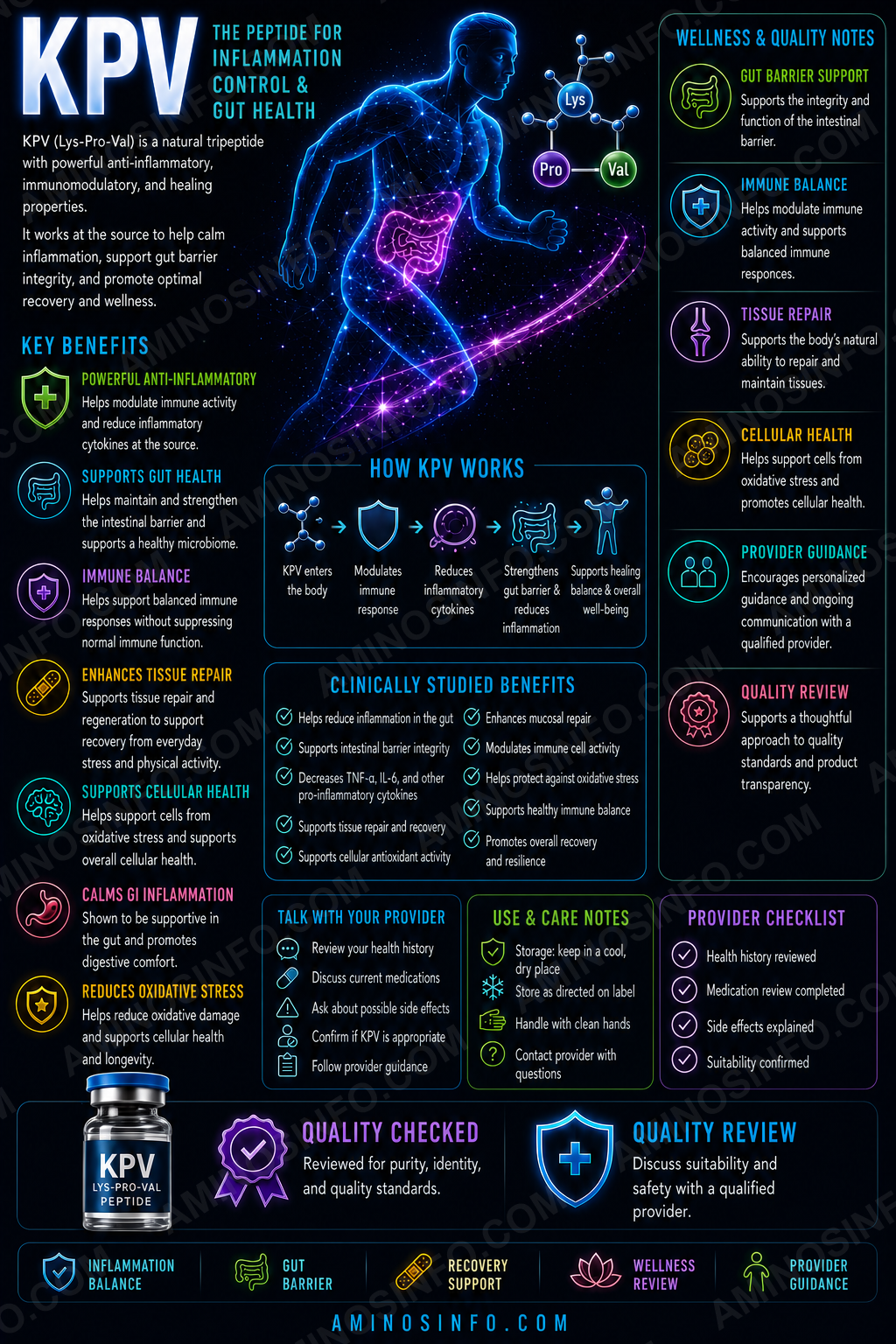
A comprehensive, evidence-graded review of KPV, the naturally occurring Lys–Pro–Val tripeptide derived from the C-terminal region of α-melanocyte-stimulating hormone and investigated for anti-inflammatory, intestinal, epithelial, and skin-related signaling.
What Is KPV?
KPV is a naturally occurring tripeptide composed of lysine, proline, and valine. It corresponds to amino acids 11–13 of α-melanocyte-stimulating hormone and is often called α-MSH(11–13).
Lys–Pro–Val
3 amino acids
α-MSH
125672
342.43 Da
No
Main research themes
- Intestinal inflammation
- PepT1-mediated cellular uptake
- NF-κB and MAPK-related signaling
- Colon epithelial protection
- Skin and mucosal inflammation
- Wound-healing and barrier research
- Colitis-associated carcinogenesis
🧬 Structure, Sequence, and Molecular Properties
🧪 Amino-acid sequence
H-Lys-Pro-Val-OH
| Molecular formula | C16H30N4O4 |
|---|---|
| Molecular weight | 342.43 g/mol |
| CAS number | 67727-97-3 |
| PubChem CID | 125672 |
| Peptide bonds | Two |
| Disulfide bonds | None |
| Terminal form | Free N terminus and free C-terminal carboxyl group |
Salt-form differences
KPV may be supplied as free peptide, acetate salt, or TFA salt. The acetate record has a molecular weight near 402.5 Da, while a 1:1 TFA salt is approximately 456.5 Da. Gross vial weight must therefore not be confused with net KPV content.
Relationship to α-MSH
α-MSH is a 13-amino-acid melanocortin peptide involved in pigmentation, inflammation, appetite biology, and immune regulation. KPV is its C-terminal tripeptide.
Retained properties
- Anti-inflammatory signaling
- Suppression of selected cytokine pathways
- Effects on epithelial and immune cells
Reduced or absent properties
- Meaningful melanotropic pigmentation activity
- Full α-MSH receptor profile
- Broader central melanocortin effects
Several studies indicate that KPV’s anti-inflammatory effects may be partly independent of MC1R.
📅 Research Timeline
- 2003: Comparative work dissected anti-inflammatory effects of α-MSH fragments, including KPV.
- 2007–2008: PepT1-mediated uptake and murine colitis studies established KPV as a major intestinal anti-inflammatory research peptide.
- 2016: PepT1-transported KPV reduced colitis-associated tumor development in mice.
- 2017: Hyaluronic-acid-functionalized nanoparticles improved oral colon targeting and anti-colitis effects.
- 2017 onward: Microneedle, iontophoretic, nanoparticle, hydrogel, and oral targeting systems were explored.
- 2025–2026: Research increasingly focused on skin stress, wound repair, ROS-triggered oral prodrugs, and improved colon accumulation.
🧠 How Does KPV Work?
NF-κB and cytokines
KPV has been reported to suppress NF-κB activation and reduce TNF-α, IL-1β, IL-6, IL-8, and related mediators, depending on the model.
MAPK signaling
Skin and epithelial studies suggest reduced stress-related MAPK activation.
Barrier protection
Lower local cytokine activity may help preserve tight junctions, mucosal integrity, and epithelial recovery.
MC1R-independent activity
Some colitis studies found activity despite reduced or absent MC1R involvement, suggesting additional intracellular or transporter-linked mechanisms.
PepT1-Mediated Uptake
PEPT1 is a proton-coupled transporter for dipeptides and tripeptides. It is highly expressed in the small intestine and may be induced in inflamed colonic epithelial and immune cells.
Because KPV is a tripeptide, it can use PepT1 for cellular entry. PepT1-deficient models show reduced or absent KPV effects in some experiments, supporting a mechanistic role for the transporter.
Intestinal and Colitis Research
KPV reduced inflammatory signaling in intestinal epithelial and immune-cell models. In DSS and TNBS mouse colitis, it reduced weight loss, histologic injury, neutrophil activity, and inflammatory markers.
Colon-targeted nanoparticles, polysaccharide carriers, and inflammation-responsive conjugates improve local accumulation and protect KPV from gastrointestinal degradation. Some systems combined inflammation reduction with improved mucosal healing.
Clinical limitation: No large randomized human trial has established KPV as a treatment for ulcerative colitis or Crohn’s disease.
Colitis-Associated Cancer Research
In mouse models, PepT1-mediated KPV uptake reduced inflammatory signaling and colitis-associated tumor development. This supports an experimental link between inflammation control and tumor prevention, but it does not establish KPV as an anticancer drug or human colorectal-cancer preventive therapy.
Skin, Barrier, and Wound Research
KPV and related α-MSH fragments are studied for suppressing inflammatory mediators in keratinocytes and immune cells. Recent keratinocyte research suggests KPV may reduce oxidative stress, apoptosis, and MAPK/NF-κB activation after particulate exposure.
Human-skin experiments have evaluated iontophoresis, microneedles, and combined delivery because KPV is small but hydrophilic. Direct wound-healing evidence remains less developed than its intestinal evidence.
Delivery-System Research
- Free oral peptide: Vulnerable to degradation, dilution, and poor colon targeting.
- Hyaluronic-acid nanoparticles: Improve delivery to inflamed colonic tissue.
- ROS-responsive conjugates: Release KPV preferentially in inflamed GI environments.
- Microneedles and iontophoresis: Improve transdermal transport.
- Hydrogels and local systems: Increase local exposure while limiting systemic exposure.
Potential Side Effects and Safety Considerations
KPV’s small size and natural origin do not prove safety when concentrated, injected, repeatedly administered, or delivered systemically.
- Injection-site irritation
- Hypersensitivity or immune reactions
- Unknown effects on normal immune surveillance
- Unknown reproductive and long-term toxicology
- Contamination or endotoxin in unapproved products
- Incorrect net content due to salt or water
- Unexpected effects from altered sequences or epimers
Inflammatory signaling is also necessary for infection control and tissue repair; excessive suppression could theoretically impair host defense.
🧪 Laboratory Testing Methods
| Method | Purpose | Limitation |
|---|---|---|
| RP-HPLC / UPLC | Separates KPV from deletion products and synthesis impurities | Area purity does not prove sequence or net content |
| LC-MS / HRMS | Confirms parent mass near 342.43 Da | Sequence isomers can share formula and mass |
| MS/MS sequencing | Confirms Lys–Pro–Val residue order | Short peptides require optimized fragmentation |
| Chiral analysis | Confirms L stereochemistry and detects epimers | Hydrolysis may introduce racemization |
| Net peptide-content assay | Measures actual KPV mass | Must correct for water, acetate, TFA, and salts |
| PepT1 uptake assay | Measures transporter-dependent entry | Transporter expression differs by model |
| NF-κB reporter and cytokine assays | Measure anti-inflammatory potency | Response is cell- and stimulus-dependent |
| Barrier assays | Measure TEER, permeability, and junction proteins | In-vitro barriers do not reproduce whole intestine |
| Protease-stability assay | Measures GI, plasma, or skin degradation | May not predict human exposure |
| Endotoxin, sterility, bioburden | Required for relevant formulations | Does not establish approved human safety |
📄 How to Interpret a KPV COA
- Verify the exact sequence H-Lys-Pro-Val-OH.
- Do not accept intact mass alone; sequence isomers can share mass.
- Use MS/MS to confirm K–P–V order.
- Confirm all residues are L-configured.
- Confirm parent mass near 342.43 Da.
- State free peptide, acetate, or TFA salt.
- Report net peptide content after correcting for water and counterions.
- Measure deletion products, epimers, and residual synthesis reagents.
- Use PepT1 uptake and NF-κB/cytokine assays for functional potency.
- For intestinal research, include protease stability and barrier assays.
- For parenteral research, require sterility, endotoxin, particles, and container controls.
- A COA does not prove human safety or clinical efficacy.
📊 Comparison Tables
KPV vs α-MSH vs KdPT vs Selank
| Feature | KPV | α-MSH | KdPT | Selank |
|---|---|---|---|---|
| Length | 3 aa | 13 aa | 3 aa | 7 aa |
| Main focus | Gut/skin inflammation | Melanocortin and pigmentation biology | Anti-inflammatory research | Neuroimmune research |
| Pigmentation | Minimal or absent | Present | Minimal | Not primary |
| FDA approved | No | No as native peptide drug | No | No |
KPV vs BPC-157 vs Larazotide vs LL-37
| Feature | KPV | BPC-157 | Larazotide | LL-37 |
|---|---|---|---|---|
| Main focus | Inflammatory signaling | Tissue-protection research | Tight-junction regulation | Host defense |
| Gut mechanism | PepT1, NF-κB | Cytoprotective pathways | Barrier-junction regulation | Antimicrobial and immune signaling |
| Human trials | Very limited | No major formal trials | Yes | Limited |
| FDA approved | No | No | No | No |
Free KPV vs Colon-Targeted KPV
| Feature | Free KPV | Targeted nanoparticle/prodrug |
|---|---|---|
| GI stability | Limited | Improved |
| Colon accumulation | Low or variable | Higher in animal models |
| Inflammation targeting | Depends on local transport | Carrier and ROS/CD44 targeting |
| Development complexity | Low | High |
🖼️ Original Diagram Specifications
- Peptide architecture: Lys–Pro–Val with termini labeled.
- α-MSH origin: Highlight residues 11–13.
- PepT1 mechanism: Inflamed colon, transporter uptake, intracellular signaling.
- NF-κB pathway: Reduced activation and cytokine output.
- Colitis model: Barrier injury, KPV uptake, reduced inflammation, mucosal repair.
- Delivery systems: Free peptide, nanoparticle, ROS-triggered conjugate, microneedle.
- COA workflow: Sequence, chirality, mass, salt correction, potency, microbiology.
❓ Frequently Asked Questions
Is KPV a peptide?
Yes, a Lys–Pro–Val tripeptide.
What is its formula?
C₁₆H₃₀N₄O₄.
What is its molecular weight?
342.43 Da for the free peptide.
What is the CAS number?
67727-97-3.
Is KPV FDA approved?
No.
Does it cause tanning?
No meaningful pigment-inducing activity has been established.
What is it studied for?
Intestinal inflammation, PepT1 transport, epithelial barriers, skin inflammation, and targeted delivery.
Does it treat ulcerative colitis?
Animal results are promising, but human efficacy is unproven.
Can it be absorbed orally?
It can use PepT1, but free oral delivery faces degradation and targeting limits.
Is there an established injectable dose?
No.
Does 99% HPLC purity prove identity?
No. Sequence order, chirality, salt form, net content, and functional potency must also be confirmed.
Final Thoughts
KPV is one of the smallest biologically active melanocortin-derived peptides. Despite containing only three amino acids, it has shown reproducible anti-inflammatory effects in intestinal epithelial cells, immune cells, and animal models of colitis.
Its most distinctive mechanism is transport through PepT1, which can become elevated in inflamed colonic tissue. Downstream effects include reduced NF-κB activation, lower cytokine production, and improved epithelial-barrier conditions.
The strongest evidence remains preclinical. Colon-targeted nanoparticles and inflammation-responsive prodrugs improve delivery in mice, but no large human trial has established KPV as a treatment for inflammatory bowel disease, skin disease, wounds, or systemic inflammation.
Analytical authentication requires sequence confirmation by MS/MS, L stereochemistry, terminal structure, salt form, net peptide content, impurities, PepT1 transport, anti-inflammatory potency, stability, and microbiological quality.
📚 References
- Dalmasso G, et al. PepT1-Mediated Tripeptide KPV Uptake Reduces Intestinal Inflammation. Gastroenterology. 2008.
- Kannengiesser K, et al. Melanocortin-Derived Tripeptide KPV in Murine Inflammatory Bowel Disease. Inflammatory Bowel Diseases. 2008.
- Getting SJ, et al. Dissection of the Anti-inflammatory Effect of α-MSH Fragments. Journal of Pharmacology and Experimental Therapeutics. 2003.
- Luger TA, et al. α-MSH-Related Peptides as Anti-inflammatory Agents. Annals of the Rheumatic Diseases. 2007.
- Böhm M, et al. Melanocortin Peptides and Cutaneous Wound Healing. Experimental Dermatology. 2019.
- Xiao B, et al. Orally Targeted KPV Nanoparticles for Ulcerative Colitis. Molecular Therapy. 2017.
- Viennois E, et al. PepT1, KPV, and Colitis-Associated Cancer. Cancer Research. 2016.
- Bettenworth D, et al. KdPT and Intestinal Barrier Function. American Journal of Pathology. 2011.
- Pawar K, et al. Transdermal Iontophoretic Delivery of KPV. Drug Delivery and Translational Research. 2017.
- Cheng J, et al. Inflammation-Triggered Oral Anti-inflammatory Peptide Delivery. 2026.
- Songok AC, et al. Structural Modification of KPV. 2018.
- Adnan SB, et al. Tripeptides in Wound Healing and Skin Regeneration. 2025.
- PubChem. MSH(11-13), CID 125672.
- PubChem. L-Lysyl-L-Prolyl-L-Valine Acetate.
- Brzoska T, et al. α-MSH and Related Tripeptides. Endocrine Reviews. 2008.
- Catania A, et al. Melanocortins: Multiple Actions and Therapeutic Potential. Pharmacological Reviews.
- ICH Q1A(R2), Q2(R2), Q3A, Q3B, Q3C, and Q6B.
- USP <621>, <71>, <85>, and <788>.
Chemistry, α-MSH origin, PepT1 transport, intestinal, skin, delivery-system, safety, regulatory, and analytical information reviewed in July 2026.