Thymalin: What It Is, How It Works, Benefits, and Research Overview
A detailed, evidence-graded review of Thymalin, including its identity as a bovine thymus-derived polypeptide complex, proposed active constituents KE, EW, and EDP, immune and hematopoietic research, aging studies, inflammatory and gene-regulation hypotheses, safety, analytical testing, and COA interpretation.
What Is Thymalin?
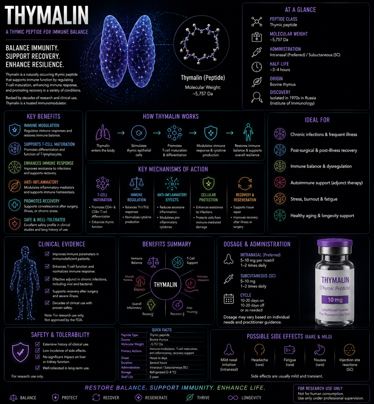
Thymalin is a complex preparation of low-molecular-weight peptides isolated from animal thymus tissue, traditionally the thymus of young calves. It was developed in the Soviet and Russian peptide-bioregulator research programs associated with Vladimir Khavinson and colleagues.
Unlike Thymogen, Vilon, Crystagen, Epitalon, or Thymosin Alpha-1, Thymalin is not a single chemically defined sequence. Its biological activity is attributed to a mixture of short peptides and possibly other low-molecular-weight constituents produced during extraction and purification.
Thymus-derived peptide complex
Calf thymus
No
KE, EW, EDP
Immune and hematopoietic regulation
No
🧬 Composition, Structure, and Molecular Identity
No single molecular structure
Because Thymalin is a peptide mixture, it cannot be represented by one structural formula, one exact mass, or one sequence.
Proposed active constituents
Recent Khavinson-group reviews identify several short peptides as biologically relevant components or functional constituents of Thymalin:
| Peptide | Sequence | Common name or role | Approximate molecular weight |
|---|---|---|---|
| KE | Lys–Glu | Vilon; immune and gene-regulation research | 275.3 g/mol |
| EW | Glu–Trp | Thymogen / oglufanide; immune and hematopoietic research | 333.3 g/mol |
| EDP | Glu–Asp–Pro | Crystagen-associated thymic peptide | 359.3 g/mol |
Mixture complexity
These three peptides do not necessarily account for the full composition or biological activity of every Thymalin preparation. Extraction source, animal age, tissue handling, enzymatic degradation, purification, sterilization, and storage can affect the final peptide fingerprint.
⚛️ Molecular Weight and 🧫 Formula
| Single molecular formula | Not applicable |
|---|---|
| Single molecular weight | Not applicable |
| Peptide-size range | Low-molecular-weight peptide mixture; exact distribution is product dependent |
| Source material | Traditionally bovine thymus extract |
| Required identity approach | Peptide fingerprint plus quantitative marker-peptide analysis |
Statements that Thymalin is “2–8 amino acids” may be reasonable as a general description of the low-molecular-weight fraction, but the full composition is not universally standardized in public literature.
📅 Discovery Timeline and Research History
1960s–1970s: Thymic extracts studied
Scientists investigated thymus-derived factors involved in lymphocyte differentiation, immune regulation, and recovery from immune suppression.
1970s–1980s: Thymalin developed
A low-molecular-weight calf-thymus peptide preparation was developed and studied as an immunomodulatory medicine in the Soviet Union.
1980s–1990s: Clinical and experimental use expands
Research examined infection, immune deficiency, radiation and chemotherapy-associated suppression, wound healing, aging, and hematopoiesis.
1990s–2000s: Defined active fragments studied
Short peptides including EW, KE, and EDP were investigated as candidate mediators of Thymalin’s biological effects.
2002–2003: Long-term gerontology reports
Russian studies reported reduced mortality in older adults receiving repeated courses of Thymalin, Epithalamin, or both. These findings have not been independently confirmed in modern large randomized trials.
2020: Human hematopoietic stem-cell study
Thymalin reduced CD44 and CD117 expression and increased CD28 expression in cultured human hematopoietic stem cells, which investigators interpreted as accelerated differentiation toward mature T-cell phenotypes.
2021–2023: COVID-19 and molecular-mechanism publications
Research examined severe COVID-19, inflammatory signaling, DNA interactions, and gene-expression effects of KE and EW.
Current status
Thymalin remains a regional peptide medicine and research preparation without FDA approval or globally harmonized composition standards.
Thymus and Immune-System Biology
T-cell development
The thymus supports T-cell lineage commitment, positive selection, negative selection, and immune tolerance.
Thymic involution
The thymus shrinks and changes composition with age, reducing naïve T-cell output and contributing to immunosenescence.
Bone marrow–thymus connection
Hematopoietic stem cells arise in bone marrow, generate lymphoid progenitors, and supply cells that mature within the thymus.
Peripheral immune regulation
Mature T cells, antigen-presenting cells, cytokines, endocrine signals, and tissue environments determine immune function outside the thymus.
Thymic extracts are not thymus replacement
Administering thymic peptides has not been shown to rebuild a structurally youthful human thymus.
🧠 Proposed Mechanisms of Action
Thymalin’s mechanism cannot be reduced to one receptor because the product is a mixture and different constituents may act through different pathways.
1. Hematopoietic differentiation
A human cell-culture study reported reduced stem and intermediate markers CD44 and CD117 with increased CD28, consistent with differentiation toward mature T-cell-associated phenotypes.
2. T-cell regulation
Thymalin has been studied for effects on T-cell number, function, differentiation, and immune coordination.
3. Cytokine modulation
Thymalin and its proposed components have been associated with changes in inflammatory mediators such as TNF and IL-6 in cell models.
4. Gene-expression regulation
KE, EW, and EDP have been proposed to enter the nucleus or nucleolus and influence promoter regions, DNA interactions, chromatin, or transcription-related processes.
5. Tissue repair and stress adaptation
Regional research describes effects on regeneration, coagulation, oxidative stress, and recovery after injury or immunosuppression.
🎯 Target and Pathway Profile
| Target or pathway | Evidence status |
|---|---|
| CD44 | Reduced in cultured human hematopoietic stem cells. |
| CD117 | Reduced in cultured human hematopoietic stem cells. |
| CD28 | Increased in cultured cells; interpreted as mature T-cell differentiation. |
| T-cell differentiation | Central historical and experimental research theme. |
| TNF and IL-6 | Modulated in selected cell studies involving thymic peptides. |
| DNA or chromatin interaction | Proposed for KE, EW, and EDP; physiological specificity remains uncertain. |
| Single exclusive receptor | Not applicable or established. |
T-Cell and Immune Research
T-cell differentiation
Thymalin was developed around the hypothesis that thymus-derived peptides can restore or normalize T-lymphocyte maturation and function.
CD28 signaling
CD28 is a costimulatory receptor important for T-cell activation, survival, and antiviral responses. Increased CD28 expression in vitro does not prove improved immunity in patients.
Immune deficiency and infection
Regional clinical literature describes use in secondary immune deficiency, bacterial and viral infection, postoperative recovery, and chronic disease.
Immune normalization versus stimulation
Thymalin is often described as an immunomodulator rather than a simple stimulant because excessive activation may be harmful.
No universal immune benefit
There is no large modern trial demonstrating improved infection resistance or immune performance in all healthy adults.
Hematopoietic Stem-Cell and Recovery Research
2020 human HSC study
Investigators exposed cultured human hematopoietic stem cells to Thymalin and measured differentiation markers.
Reported changes
- CD44 expression decreased approximately two- to threefold.
- CD117 expression decreased approximately two- to threefold.
- CD28 expression increased approximately 6.8-fold.
Interpretation
The authors interpreted the pattern as accelerated differentiation toward mature T-cell-associated phenotypes.
Limitations
The study did not demonstrate improved blood counts, infection survival, vaccine response, or immune recovery in a randomized patient trial.
Not a replacement for approved hematopoietic therapy
Thymalin is not an FDA-approved alternative to G-CSF, erythropoietin, stem-cell transplantation, transfusion, antibiotics, or oncology-directed supportive care.
Inflammation and Cytokine Research
Monocyte/macrophage models
Thymalin and several short thymic peptides have been tested in THP-1 cells, a leukemia-derived monocyte/macrophage model.
TNF and IL-6 responses
Peptide exposure altered inflammatory responses after lipopolysaccharide stimulation, suggesting priming or tolerance-like effects.
Mixture attribution problem
A response to Thymalin cannot automatically be attributed to KE, EW, or EDP individually unless each peptide is tested separately.
Inflammatory balance is context dependent
Reducing cytokine release may protect tissue in one setting but impair pathogen clearance in another.
No established anti-inflammatory indication
Thymalin is not proven to treat rheumatoid arthritis, inflammatory bowel disease, asthma, vasculitis, sepsis, or cytokine storm.
Aging, Immunosenescence, and Longevity Claims
Immunosenescence
Aging affects thymic output, T-cell diversity, bone-marrow function, inflammatory signaling, vaccine response, and immune memory.
Long-term Russian studies
Studies published in the early 2000s reported lower mortality among older adults receiving repeated courses of Thymalin, Epithalamin, or both.
Why caution is required
- Study methods are difficult to compare with modern randomized trial standards.
- Blinding and allocation methods are not always clear.
- Multiple peptide preparations were sometimes used.
- Independent replication is lacking.
- Changes in background medical care may confound long-term results.
No proven human lifespan extension
The existing literature does not establish that Thymalin extends human life or reverses biological aging.
No proven thymic rejuvenation
Improved immune markers do not prove anatomical regrowth or restoration of a youthful thymus.
Infection and COVID-19 Research
Historical infection studies
Regional literature describes use in bacterial and viral infection, immune suppression, and postoperative complications.
Severe COVID-19 studies
Small Russian studies reported improvements in selected immune markers and outcomes when Thymalin was added to standard therapy in older patients.
Important limitations
Many studies were small, single-center, open-label, or conducted during rapidly changing standards of COVID-19 treatment.
Not an antiviral
Thymalin does not directly replace antiviral drugs, vaccination, oxygen, corticosteroids, anticoagulation, antibiotics, or critical care.
COVID-era findings are not general immune proof
Results from one infection cannot be generalized to influenza, HIV, hepatitis, bacterial sepsis, or healthy-aging use.
Gene-Expression and Chromatin Hypotheses
Short-peptide theory
Khavinson-school research proposes that ultrashort peptides can enter cells and interact with DNA, histones, promoter regions, or transcription factors.
KE and EW modeling
Molecular-modeling studies have examined interactions of KE and EW with double-stranded DNA.
Gene and protein expression
Cell studies report changes in immune-related genes and proteins after exposure to Thymalin-associated peptides.
Binding does not prove regulation
Docking, molecular modeling, or in vitro DNA interaction does not establish selective gene regulation in living humans.
Mixture complexity
Batch-to-batch differences may alter which peptide components reach cells and at what concentrations.
Evidence Limitations and Clinical Interpretation
Product heterogeneity
Extract-based peptide mixtures may vary by source tissue, purification, manufacturing, and storage.
Regional evidence concentration
Much of the literature comes from a limited network of Russian institutions and investigators.
Older trial standards
Many studies predate current expectations for registration, blinding, allocation concealment, pharmacokinetics, and reproducible product characterization.
Defined-peptide evidence cannot fully validate the mixture
KE, EW, and EDP studies help generate hypotheses but do not prove that every Thymalin batch contains effective or equivalent amounts.
Biomarkers are not clinical outcomes
Changes in CD markers, cytokines, or gene expression do not automatically prove fewer infections, longer survival, or improved quality of life.
Safety and Regulatory Considerations
No standardized US prescribing information
No FDA-approved label defines dose, route, pharmacokinetics, contraindications, interactions, pregnancy safety, or long-term adverse effects.
Animal-derived material
Thymus extracts require controls for species identity, transmissible agents, adventitious viruses, prions, bacteria, endotoxin, and residual tissue proteins.
Immune modulation
Potential effects on T cells and cytokines may be relevant in autoimmune disease, transplantation, immunosuppressive therapy, and cancer.
Allergy and hypersensitivity
Animal-derived peptide mixtures may carry higher immunogenicity and allergy concerns than one fully synthetic defined peptide.
Product-quality risk
Unapproved products may be mislabeled, undercharacterized, contaminated, or substituted with one synthetic peptide while being sold as the full complex.
Regulatory status
Thymalin is not FDA approved in the United States.
🧪 Laboratory Testing Methods
| Method | Purpose | Important limitation |
|---|---|---|
| LC-MS peptide fingerprinting | Defines the mixture’s peptide profile and detects batch variation. | No single peak can establish full identity. |
| Targeted LC-MS/MS | Confirms and quantifies KE, EW, EDP, and other marker peptides. | Marker peptides may not capture all active components. |
| Size-exclusion chromatography | Characterizes molecular-size distribution. | Low resolution for very small peptides. |
| RP-HPLC or UPLC | Provides chromatographic fingerprint and impurity profile. | Area percentages cannot be interpreted like purity of one compound. |
| Amino-acid analysis | Measures overall amino-acid composition. | Does not identify peptide sequences. |
| Total peptide or nitrogen assay | Measures total peptide content. | Does not establish active composition. |
| Species-identity testing | Confirms bovine source and detects adulteration. | Requires validated DNA or protein methods. |
| Residual host-protein testing | Measures larger bovine proteins or tissue contaminants. | Acceptable limits depend on manufacturing and route. |
| Viral and adventitious-agent testing | Evaluates animal-source biological safety. | Must be integrated with sourcing and manufacturing controls. |
| Prion/TSE risk assessment | Assesses transmissible spongiform encephalopathy risk. | Testing alone cannot replace controlled source-country and tissue selection. |
| Cell-based potency assay | May assess HSC differentiation, CD28, cytokines, or T-cell responses. | No universally accepted Thymalin potency assay exists. |
| Sterility, endotoxin, and particles | Required for injectable final products. | Raw-powder results do not certify the finished dosage form. |
| Residual solvents and elemental impurities | Evaluates manufacturing contaminants. | Does not prove biological equivalence. |
| Stability-indicating fingerprint | Tracks peptide degradation and batch-profile change over time. | Requires predefined acceptance ranges for a complex mixture. |
📄 How to Interpret a Thymalin COA
- Reject a single-sequence COA: Authentic Thymalin is a mixture, not one peptide.
- Require a chromatographic and mass-spectrometric fingerprint: The batch should match a qualified reference standard.
- Confirm marker peptides: KE, EW, and EDP should be identified and preferably quantified if claimed as active constituents.
- Check total peptide content: This must be distinct from HPLC “purity.”
- Review molecular-size distribution: Larger proteins and uncharacterized high-molecular-weight material should be controlled.
- Verify animal source: Species, tissue source, country of origin, health status, and traceability matter.
- Review viral, microbial, endotoxin, and TSE controls: These are critical for animal-derived material.
- Check batch-to-batch comparability: One passing lot does not validate future lots without fingerprint matching.
- Match testing to the final route: Injectable products require final-product sterility, endotoxin, particles, and container-closure testing.
- Do not infer efficacy: A COA cannot prove immune restoration, hematopoietic recovery, anti-aging effects, lower infection risk, or longer life.
📊 Thymalin vs Thymogen vs Vilon vs Epitalon
| Feature | Thymalin | Thymogen | Vilon | Epitalon |
|---|---|---|---|---|
| Composition | Thymus-derived peptide mixture | EW dipeptide | KE dipeptide | AEDG tetrapeptide |
| Single defined sequence? | No | Yes | Yes | Yes |
| Main research focus | Broad thymic immune regulation | Immune and hematopoietic signaling | Immune and gene regulation | Pineal, circadian, and telomere research |
| Animal-derived? | Traditionally yes | No | No | No |
| FDA approved? | No | No | No | No |
Thymalin vs Thymosin Alpha-1 vs Thymulin
| Feature | Thymalin | Thymosin Alpha-1 | Thymulin |
|---|---|---|---|
| Structure | Peptide mixture | 28-AA acetylated peptide | Zinc-dependent nonapeptide |
| Main biology | Broad thymic extract effects | Innate and adaptive immune modulation | Immune–neuroendocrine signaling |
| Composition control | Complex fingerprint required | Single sequence | Single peptide plus zinc complex |
| Human clinical evidence | Regional and heterogeneous | Substantial international literature | Mainly mechanistic and preclinical |
Thymalin vs Defined Synthetic Blend
| Feature | Animal-derived Thymalin | KE + EW + EDP synthetic blend |
|---|---|---|
| Composition | Complex and partly undefined | Three defined peptides |
| Biological equivalence | Original reference preparation | Not established automatically |
| Source risks | Animal-source contaminants and variability | Synthetic impurities and blend-ratio errors |
| COA approach | Fingerprint plus marker quantification | Identity and amount of each peptide |
Thymalin vs Evidence-Based Immune Recovery
| Approach | Established role | Difference from Thymalin |
|---|---|---|
| Vaccination | Antigen-specific immune memory | Defined preventive intervention |
| G-CSF | Neutrophil recovery after selected treatments | Approved receptor-targeted growth factor |
| Antimicrobials | Direct pathogen treatment | Pathogen-specific therapy |
| Stem-cell transplantation | Reconstitutes hematopoiesis in selected diseases | Established specialist treatment |
| Thymalin | Regional immune-modulating peptide extract | No FDA approval and less definitive evidence |
🔗 Related Peptides and Pathways
- Thymogen: EW dipeptide proposed as an active Thymalin constituent.
- Vilon: KE dipeptide associated with immune and gene-regulation research.
- Crystagen: EDP tripeptide associated with thymic differentiation research.
- Thymosin Alpha-1: Distinct defined 28-amino-acid peptide.
- Thymulin: Distinct zinc-dependent thymic hormone.
- CD28: T-cell costimulatory receptor increased in a Thymalin HSC study.
- CD44 and CD117: Stem and progenitor markers reduced in the same cell-culture study.
🖼️ Original Diagram Specifications
Diagram 1: Thymalin mixture map
Show an animal thymus extract containing multiple short peptides, highlighting KE, EW, EDP, and additional unidentified low-molecular-weight components.
Diagram 2: Thymalin versus defined peptides
Contrast the complex extract with single-molecule structures for Thymogen, Vilon, Crystagen, and Thymosin Alpha-1.
Diagram 3: Bone marrow–thymus pathway
Show hematopoietic stem cells, lymphoid progenitors, thymic maturation, CD4/CD8 T cells, and peripheral immune function.
Diagram 4: HSC marker experiment
Show Thymalin exposure associated with lower CD44/CD117 and higher CD28, with all findings labeled in vitro.
Diagram 5: Aging and immunosenescence
Show thymic involution, lower naïve T-cell output, reduced repertoire diversity, inflammaging, and infection vulnerability.
Diagram 6: Evidence ladder
Show peptide fingerprinting, cell culture, regional clinical studies, independently replicated randomized trials, regulatory approval, and confirmed mortality benefit.
Diagram 7: COA workflow
Show species verification, LC-MS fingerprint, marker-peptide quantification, total peptide content, size distribution, host proteins, viral and TSE risk controls, sterility, endotoxin, and stability.
❓ Frequently Asked Questions
Is Thymalin a peptide?
It is more accurately described as a complex mixture of low-molecular-weight thymus-derived peptides.
What is Thymalin’s amino-acid sequence?
It has no single sequence.
What is its molecular weight?
There is no single molecular weight because it is a mixture.
What peptides are found in Thymalin?
KE, EW, and EDP are frequently described as biologically relevant components, but the complete mixture is not limited to these sequences.
Is Thymalin the same as Thymogen?
No. Thymogen is the defined EW dipeptide; Thymalin is a broader peptide extract.
Is Thymalin the same as Thymosin Alpha-1?
No. Thymosin Alpha-1 is a defined 28-amino-acid peptide.
Is Thymalin FDA approved?
No.
Does Thymalin increase T cells?
Cell and regional clinical research suggests effects on T-cell differentiation and immune markers, but broad modern validation is limited.
Does it support blood-cell recovery?
Experimental research suggests possible hematopoietic effects, but it is not an FDA-approved hematopoietic growth factor.
Does Thymalin reverse immune aging?
No robust evidence demonstrates reversal of immunosenescence or restoration of a youthful thymus.
Does it extend lifespan?
Older Russian studies reported lower mortality, but these findings have not been confirmed by large independent modern trials.
Can a synthetic KE/EW/EDP blend be called Thymalin?
Not automatically. Biological and analytical equivalence to the original extract must be demonstrated.
Can a COA report “99% purity” for Thymalin?
A single purity percentage is inadequate for a complex mixture. A validated fingerprint and marker-peptide profile are needed.
Final Thoughts
Thymalin is a biologically complex thymus-derived peptide preparation, not a single peptide. Its research history includes T-cell regulation, hematopoietic differentiation, immune recovery, infection, inflammation, and aging.
The strongest modern mechanistic evidence includes a cultured human hematopoietic stem-cell study showing reduced CD44 and CD117 with increased CD28 expression. Long-term human longevity claims and COVID-19 findings remain insufficiently independently replicated.
Legitimate Thymalin characterization requires a batch fingerprint, marker-peptide identification and quantification, total peptide content, molecular-size distribution, species and tissue traceability, host-protein controls, adventitious-agent and TSE risk assessment, route-specific microbiological testing, and stability data.
📚 References
- Khavinson VK, et al. The Use of Thymalin for Immunocorrection and Molecular Aspects of Thymic Peptides. 2021.
- Khavinson VK, et al. Thymalin: Activation of Differentiation of Human Hematopoietic Stem Cells. 2020.
- Khavinson VK, et al. PubMed record: Thymalin and human hematopoietic stem-cell differentiation. 2020.
- Kuznik B, et al. Peptide Drug Thymalin Regulates Immune Status in Severe COVID-19 Older Patients. 2021.
- Khavinson VK, et al. Results and Prospects of Using an Activator of Hematopoietic Stem Cell Differentiation in COVID-19. 2021.
- Linkova N, et al. The Influence of KE and EW Dipeptides on Gene Expression and Protein Synthesis. 2023.
- Linkova N, et al. PubMed record: KE and EW dipeptides in Thymalin-related inflammatory research. 2023.
- Avolio F, et al. Peptides Regulating Proliferative Activity and Inflammatory Pathways in THP-1 Cells. 2022.
- Khavinson VK, et al. Peptides of Pineal Gland and Thymus Prolong Human Life. 2003.
- Khavinson VK, et al. Geroprotective Effect of Thymalin and Epithalamin. 2002.
- Khavinson VK. Peptides and Ageing. 2002.
- Anisimov VN, Khavinson VK. Peptide Bioregulation of Aging: Results and Prospects. Biogerontology. 2010.
- Wolf E, et al. Thymic Peptides for Treatment of Cancer Patients. Cochrane review.
- Shevyrev D, et al. Hematopoietic Stem Cells and the Immune System. 2023.
- Seita J, Weissman IL. Hematopoietic Stem Cell: Self-Renewal versus Differentiation. 2010.
- Goyani P, et al. Immunosenescence: Aging and Immune System Decline. 2024.
- Palmer DB. The effect of age on thymic function. Frontiers in Immunology.
- Thomas R, Wang W, Su DM. Age-related thymic involution and immunosenescence. Immunity & Ageing.
- Nikolich-Žugich J. The twilight of immunity. Nature Immunology.
- Goronzy JJ, Weyand CM. Immune aging and vaccine responses. Nature Immunology.
- Murphy K, Weaver C. Janeway's Immunobiology. Garland Science.
- Kaech SM, Wherry EJ, Ahmed R. Effector and memory T-cell differentiation. Nature Reviews Immunology.
- Sharpe AH, Freeman GJ. The B7-CD28 superfamily. Nature Reviews Immunology.
- Chen L, Flies DB. Molecular mechanisms of T-cell co-stimulation and co-inhibition. Nature Reviews Immunology.
- Wilson A, Trumpp A. Bone-marrow stem-cell niches. Nature Reviews Immunology.
- Morrison SJ, Scadden DT. The bone-marrow niche for haematopoietic stem cells. Nature.
- Orkin SH, Zon LI. Hematopoiesis: an evolving paradigm for stem-cell biology. Cell.
- Turner MD, et al. Cytokines and inflammatory regulation. Biochimica et Biophysica Acta.
- Lawrence T. The NF-κB pathway in inflammation. Cold Spring Harbor Perspectives in Biology.
- Murray PJ. Macrophage activation and polarization. Annual Review of Physiology.
- International Council for Harmonisation. ICH Q2(R2): Validation of Analytical Procedures.
- United States Pharmacopeia. General Chapter <621>, Chromatography.
- United States Pharmacopeia. General Chapter <71>, Sterility Tests.
- United States Pharmacopeia. General Chapter <85>, Bacterial Endotoxins Test.
- United States Pharmacopeia. General Chapters <232> and <233>, Elemental Impurities.
- International Council for Harmonisation. ICH Q3C: Residual Solvents.
- International Council for Harmonisation. ICH Q1A(R2): Stability Testing of New Drug Substances and Products.
- ICH Q5A. Viral Safety Evaluation of Biotechnology Products.
- ICH Q5D. Derivation and Characterisation of Cell Substrates Used for Production.
- World Health Organization. Guidelines on transmissible spongiform encephalopathy risk and animal-derived medicinal materials.
- European Medicines Agency. Note for guidance on minimizing the risk of transmitting animal spongiform encephalopathy agents via medicinal products.
Identity, composition, hematopoietic, immune, aging, infection, safety, and analytical findings were reviewed in July 2026. Thymalin remains unapproved by the FDA in the United States.