Retinalamin: What It Is, How It Works, Benefits, and Research Overview
A corrected, evidence-graded review of Retinalamin, including its identity as a cattle-retina-derived polypeptide complex, retinal mechanisms, glaucoma and diabetic-retinopathy studies, photochemical-injury research, safety, testing, and COA interpretation.
What Is Retinalamin?
Retinalamin is a low-molecular-weight polypeptide preparation extracted from cattle retinal tissue. It is manufactured as a lyophilized prescription product for intramuscular or parabulbar administration in Russia and selected regional markets.
Research focuses on retinal neurons, photoreceptors, retinal pigment epithelium, Müller glia, retinal ganglion cells, vascular permeability, inflammation, oxidative stress, and excitotoxic injury.
Retinalamin
Retinal polypeptide complex
Cattle retina
No
Retinal protection
No
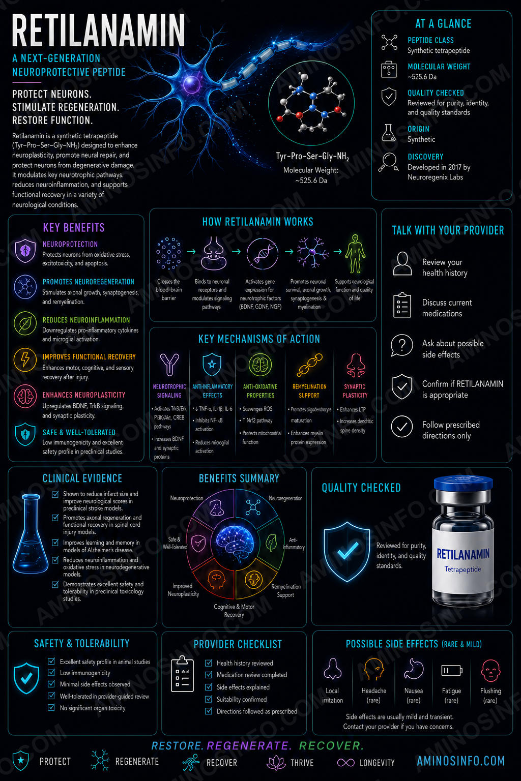
Scientific Corrections to the Original Draft
| Original claim | Correction |
|---|---|
| Retilanamin is a defined neuroactive peptide. | No recognized compound by that name was identified; the likely intended product is Retinalamin. |
| Designed for cognition and memory. | Retinalamin is studied in ophthalmology, not as a general nootropic. |
| Promotes dendritic spines and LTP. | No direct evidence establishes cortical synaptogenesis or long-term-potentiation enhancement. |
| Mimics BDNF or CNTF. | No single neurotrophic receptor or BDNF/CNTF-mimetic mechanism has been established. |
| Single synthetic sequence. | It is a heterogeneous animal-derived polypeptide fraction. |
| Used in Alzheimer’s or Parkinson’s models. | The identifiable literature concerns retina and optic nerve. |
🧬 Composition, Structure, Sequence, Formula, and Molecular Weight
Retinalamin is a peptide mixture, not one chemical entity.
🧪 Sequence
No single amino-acid sequence applies. A product represented by one short sequence, such as Glu–Lys or Lys–Glu, should not automatically be accepted as authentic Retinalamin.
| Single molecular formula | Not applicable |
|---|---|
| Single molecular weight | Not applicable |
| Reported fraction range | Water-soluble polypeptides below approximately 10 kDa |
| Source | Cattle retinal tissue |
| Official active-substance description | Cattle retinal polypeptides |
| Typical marketed vial | 5 mg retinal polypeptide complex plus 17 mg glycine stabilizer |
Some research-product sellers list a single dipeptide, formula, and CAS number under the Retinalamin name. That conflicts with the official product identity and may represent a mislabeled or different synthetic product.
📅 Discovery Timeline and Research History
Late Soviet peptide-bioregulator era
Retina-derived peptide fractions were developed within Russian tissue-specific peptide programs.
1990s–2000s
Regional reports evaluated glaucoma, retinal dystrophy, diabetic retinopathy, myopia, inflammatory retinal disease, and optic neuropathy.
2003
Retinalamin and Epitalon were tested in retinal and pigment-epithelial cell cultures; concentration-dependent proliferation was reported.
2005
A small pediatric report evaluated inherited retinal degeneration.
2019
Retinalamin was reported to reduce glutamate-associated toxicity in isolated retinal-cell cultures.
2021
Functional and morphological outcomes were studied in a rabbit photochemical retinal-injury model.
2024
A newer clinical report evaluated retinal structure and function in diabetic retinopathy.
Retinal Biology and Disease Context
Photoreceptors
Rods and cones convert light into neural signals and depend on the retinal pigment epithelium and Müller cells.
Retinal pigment epithelium
The RPE recycles visual pigments, supports the blood-retina barrier, and phagocytoses photoreceptor outer segments.
Retinal ganglion cells
Their axons form the optic nerve; degeneration is central to glaucoma.
Müller glia
These cells regulate extracellular ions, glutamate clearance, and retinal metabolic support.
🧠 Proposed Mechanisms of Action
Because Retinalamin is a mixture, no single receptor or pathway explains all reported effects.
1. Retinal-cell metabolic support
Official information describes activation of ocular-tissue metabolism and intracellular protein synthesis.
2. Glutamate inactivation and Müller-cell function
The manufacturer proposes improved Müller-cell activity and glutamate inactivation.
3. Membrane and energy processes
Retinalamin is claimed to normalize membrane function and optimize cellular energy metabolism.
4. Oxidative-stress regulation
Research and labeling describe effects on lipid peroxidation and oxidative injury.
5. Vascular permeability and inflammation
Regional information describes normalization of retinal vascular permeability and reduced local inflammation.
🎯 Target and Pathway Profile
| Target/pathway | Evidence status |
|---|---|
| Retinal ganglion-cell survival | Supported in isolated-cell and glaucoma-related research. |
| Glutamate excitotoxicity | Reduced toxicity reported in retinal-cell culture. |
| Retinal pigment epithelium | Proliferative and functional claims reported. |
| Müller glia | Mechanistic claim; detailed direct evidence remains limited. |
| Oxidative stress | Supported by regional mechanistic and injury-model research. |
| Single receptor | None established. |
Major Research Areas
Glutamate excitotoxicity
A 2019 study reported that Retinalamin was not cytotoxic and reduced glutamate-associated injury in isolated retinal cells. Cell-culture protection does not prove prevention of human vision loss.
Photochemical retinal injury
A 2021 rabbit study reported protective functional and morphological findings after experimental light damage. This model does not directly establish efficacy in macular degeneration or inherited disease.
Glaucoma
Regional studies evaluate Retinalamin as an adjunct after intraocular pressure is controlled. It does not replace pressure-lowering medication, laser, surgery, or monitoring.
Diabetic retinopathy
Studies have evaluated visual function, electrophysiology, imaging, and retinal structure. It does not replace glucose and blood-pressure control, anti-VEGF therapy, laser, or vitrectomy.
Inherited retinal dystrophy
A small pediatric study reported visual changes, but mixed diagnoses, limited controls, and lack of modern genetic stratification restrict interpretation.
Regional administration
The registered product is prepared for intramuscular or parabulbar injection. Parabulbar administration requires trained ophthalmic clinicians because of procedural risks.
Evidence Limitations
- Animal-derived mixture with potential batch variability
- Research concentrated in Russia and nearby regions
- Many studies predate modern registration and masking standards
- Mixed diseases, routes, and outcome measures
- Limited independent multicenter replication
- Biomarker or electrophysiology changes do not necessarily equal durable vision preservation
Safety and Regulatory Considerations
Official regional information notes possible allergic reactions in people with individual intolerance. Parabulbar and intramuscular administration add procedural risks.
Animal-derived production requires source traceability, species verification, viral-safety controls, bacterial and endotoxin testing, residual host-protein limits, and transmissible-spongiform-encephalopathy risk management.
Retinalamin is not FDA approved in the United States.
🧪 Laboratory Testing Methods
| Method | Purpose | Limitation |
|---|---|---|
| LC-MS peptide fingerprinting | Defines mixture profile and batch consistency. | No single peak proves full identity. |
| Targeted LC-MS/MS | Identifies reproducible marker peptides. | No universally accepted public marker panel. |
| Size-exclusion chromatography | Confirms low-molecular-weight distribution. | Limited resolution for complex small peptides. |
| RP-HPLC/UPLC fingerprint | Compares chromatographic profiles. | One “purity percentage” is inappropriate for a mixture. |
| Total peptide assay | Measures active-complex amount. | Does not establish composition or potency. |
| Species-identity testing | Confirms cattle origin. | Requires validated DNA or proteomic methods. |
| Residual host-protein testing | Controls larger retinal proteins. | Product-specific limits are needed. |
| Viral/adventitious-agent testing | Evaluates biological safety. | Must be combined with manufacturing controls. |
| TSE risk assessment | Addresses prion risk. | Testing alone cannot replace safe sourcing. |
| Glycine assay | Confirms stabilizer content. | Must be separate from peptide content. |
| Retinal-cell potency assay | May assess glutamate injury or cell survival. | No universally accepted potency assay. |
| Sterility/endotoxin/particles | Required for injectable product. | Raw-material results do not certify final product. |
📄 How to Interpret a Retinalamin COA
- Reject a single-sequence COA: Authentic Retinalamin is a peptide mixture.
- Require source identity: Cattle retinal tissue and full traceability.
- Require LC-MS and chromatographic fingerprints.
- Confirm molecular-size distribution.
- Separate active peptide content from glycine stabilizer.
- Review residual host proteins, viruses, microbes, endotoxin, and TSE controls.
- Check batch-to-batch comparability.
- Require final-product sterility, particles, reconstitution, and stability testing.
- Do not infer efficacy: A COA cannot prove retinal regeneration or prevention of vision loss.
📊 Comparison Tables
Retinalamin vs Retinylamine vs Retinol
| Feature | Retinalamin | Retinylamine | Retinol |
|---|---|---|---|
| Type | Cattle-retina polypeptide complex | Small-molecule visual-cycle modulator | Vitamin A alcohol |
| Main focus | Retinal protection | Toxic retinaldehyde reduction | Vitamin A physiology |
| Single molecule? | No | Yes | Yes |
Retinalamin vs Cortexin vs Thymalin
| Feature | Retinalamin | Cortexin | Thymalin |
|---|---|---|---|
| Source tissue | Cattle retina | Animal cerebral cortex | Calf thymus |
| Main regional use | Retinal disease | Neurologic conditions | Immune regulation |
| Single sequence? | No | No | No |
Retinalamin vs Standard Eye Treatments
| Approach | Established role | Difference |
|---|---|---|
| Anti-VEGF therapy | Macular edema and neovascular disease | Defined target and modern trial evidence |
| Pressure-lowering therapy | Glaucoma | Established standard of care |
| Laser/surgery | Selected retinal and glaucoma indications | Procedure-specific evidence |
| Retinalamin | Regional peptide adjunct | Complex mixture with limited international validation |
Authentic Retinalamin vs Single Synthetic Peptide
| Feature | Authentic Retinalamin | Single peptide sold under the name |
|---|---|---|
| Composition | Complex retinal polypeptide fraction | One sequence |
| Equivalence | Reference product | Not established |
| COA approach | Fingerprint and source controls | Sequence, mass, purity, content |
🖼️ Original Diagram Specifications
- Composition map: cattle retina processed into multiple peptide fractions below 10 kDa.
- Retina cross-section: photoreceptors, RPE, Müller cells, bipolar cells, ganglion cells, vessels, optic nerve.
- Excitotoxicity pathway: glutamate, NMDA/AMPA activation, calcium, oxidative stress, apoptosis, proposed modulation.
- Disease map: glaucoma, diabetic retinopathy, retinal dystrophy, inflammation, trauma, photochemical injury.
- Administration map: parabulbar versus intramuscular delivery.
- Evidence ladder: cell culture, animal model, regional studies, multicenter trials, guideline adoption, FDA approval.
- COA workflow: source, species, fingerprint, size distribution, peptide content, glycine, host proteins, viral/TSE controls, sterility, endotoxin, particles.
❓ Frequently Asked Questions
Is “Retilanamin” recognized?
No clear compound by that name was identified. The intended name is likely Retinalamin.
Is Retinalamin a peptide?
It is a mixture of low-molecular-weight retinal polypeptides.
What is its sequence?
It has no single sequence.
What is its molecular weight?
It has no single molecular weight; the official product describes fractions below approximately 10 kDa.
Is it a cognitive-enhancement peptide?
No. Its research and regional use focus on the retina and optic nerve.
Is it FDA approved?
No.
Does it regenerate the retina?
Regional labeling uses regeneration language, but robust proof of restoring lost human retinal tissue is lacking.
Does it replace glaucoma or diabetic-retinopathy treatment?
No.
Is it the same as retinylamine?
No.
Can a single Glu–Lys peptide be called Retinalamin?
Not without equivalence evidence.
Can a COA report 99% purity?
A single purity number is inadequate for a complex extract.
Final Thoughts
The original “Retilanamin” draft described a generic cognitive neuropeptide that does not match the identifiable product. The correct compound is almost certainly Retinalamin, a cattle-retina-derived polypeptide complex used and studied regionally in ophthalmology.
Its evidence base concerns retinal ganglion cells, photoreceptors, retinal pigment epithelium, Müller glia, glutamate toxicity, oxidative injury, glaucoma, diabetic retinopathy, and retinal dystrophy—not general memory enhancement or cortical synaptic repair.
📚 References
- GEROPHARM. Official Retinalamin product information.
- Khavinson VK, et al. Effects of peptides on retinal and pigment epithelial cell proliferation. 2003.
- Suetov AA, et al. Retinoprotective effects in a rabbit photochemical-damage model. 2021.
- Avetisov SE, et al. Retinal ganglion-cell sensitivity and glutamate toxicity. 2019.
- Makashova NV, et al. Application in glaucomatous optic neuropathy. 2014.
- Strakhov VV, et al. Long-term retinal protective therapy in glaucoma. 2020.
- Khvatova AV, et al. Polypeptide bioregulators in childhood retinal abiotrophy. 2005.
- Aleksandrov EI, et al. Retinalamine in ocular tuberculosis. 2008.
- Erichev VP, et al. Peptide bioregulators: delivery and efficacy. 2020.
- Structural and functional monitoring in diabetic retinopathy. 2024.
- Pardue MT, Allen RS. Neuroprotective strategies for retinal disease. 2018.
- Parsons DE, et al. Peptidomimetics therapeutics for retinal disease. 2021.
- Yu G, et al. Peptide derivatives of retinylamine. 2021.
- Nickells RW, et al. Retinal ganglion-cell death in glaucoma. Progress in Retinal and Eye Research.
- Weinreb RN, et al. The pathophysiology and treatment of glaucoma. JAMA.
- Antonetti DA, et al. Diabetic retinopathy. New England Journal of Medicine.
- Bringmann A, et al. Müller cells in the healthy and diseased retina. Progress in Retinal and Eye Research.
- Strauss O. The retinal pigment epithelium in visual function. Physiological Reviews.
- Osborne NN, et al. Retinal ischemia and glutamate excitotoxicity. Progress in Retinal and Eye Research.
- Organisciak DT, Vaughan DK. Retinal light damage. Progress in Retinal and Eye Research.
- ICH Q2(R2). Validation of Analytical Procedures.
- ICH Q5A. Viral Safety Evaluation of Biotechnology Products.
- ICH Q3C. Residual Solvents.
- ICH Q1A(R2). Stability Testing.
- USP <621>, <71>, <85>, and <788>.
- EMA guidance on animal-derived medicinal products and TSE risk.
Identity, composition, retinal mechanisms, clinical research, safety, and analytical requirements were reviewed in July 2026. Retinalamin remains unapproved by the FDA in the United States.